


ņä¼ņ£ĀņŻ╝ļŖö ļģ╣ļé┤ņןņŚÉņä£ ļ░®ņłśņ£ĀņČ£ņØś ņĪ░ņĀłņŚÉ ņżæņÜöĒĢ£ ņŚŁĒĢĀņØä ĒĢśļŖöļŹ░, ņä¼ņ£ĀņŻ╝ņØś ļ│Ćņä▒ņØ┤ļéś ņä¼ņ£ĀņŻ╝ņäĖĒżņØś Ļ░ÉņåīļĪ£ ņØĖĒĢ┤ ņä¼ņ£ĀņŻ╝ņØś ĻĖ░ļŖźņØ┤ ņĀĆĒĢśļÉśļ®┤ ļ░®ņłśņ£ĀņČ£ļĪ£ņØś ņĀĆĒĢŁņØ┤ ņ”ØĻ░ĆļÉśņ¢┤ Ļ░£ļ░®Ļ░üļģ╣ļé┤ņןņØ┤ ņ£Āļ░£ļÉ£ļŗż.1,2 ļīĆļČĆļČäņØś ņĢłņĢĢĒĢśĻ░ĢņĀ£ļŖö ļ░®ņłś ņāØņä▒ņØä Ļ░Éņåīņŗ£ņ╝£ ņĢłņĢĢĒĢśĻ░Ģ ņ×æņÜ®ņØä ļéśĒāĆļé┤ņ¦Ćļ¦ī ĒöäļĪ£ņŖżĒāĆĻĖĆļ×Ćļöś ņĀ£ņĀ£ļŖö ĒżļÅäļ¦ēĻ│Ąļ¦ēņØä ĒåĄĒĢ£ ļ░®ņłśņ£ĀņČ£ņØä ņ”ØĻ░Ćņŗ£ņ╝£ Ļ░ĢļĀźĒĢ£ ņĢłņĢĢĒĢśĻ░Ģ ņ×æņÜ®ņØä ļéśĒāĆļé┤ņ¢┤ ļģ╣ļé┤ņןņØś ņØ╝ņ░© ņ╣śļŻīņĀ£ļĪ£ ļ¦ÄņØ┤ ņé¼ņÜ®ļÉśĻ│Ā ņ׳ļŗż.3,4 ņØ┤ņÖĆ ĒĢ©Ļ╗ś ņä¼ņ£ĀņŻ╝ļź╝ ĒåĄĒĢ£ ļ░®ņłśņ£ĀņČ£ņØä ņ”ØĻ░Ćņŗ£ĒéżļŖö ņĢĮņĀ£ņŚÉ ļīĆĒĢ┤ņä£ļÅä ļŗżņ¢æĒĢ£ ņŚ░ĻĄ¼Ļ░Ć ņŗ£Ē¢ēļÉśņ¢┤ ņÖöļŖöļŹ░5 ĻĘĖņżæ ņØ╝ņé░ĒÖöņ¦łņåī(nitric oxide, NO)Ļ░Ć ņä¼ņ£ĀņŻ╝ļź╝ ņØ┤ņÖäņŗ£ņ╝£ ļ░®ņłśņ£ĀņČ£ņØä ņ”ØĻ░Ćņŗ£Ēéżļ®┤ņä£ ņĢłņĢĢĒĢśĻ░Ģ ņ×æņÜ®ņØä ļéśĒāĆļé┤ļŖö Ļ▓āņ£╝ļĪ£ ņĢīļĀżņĀĖ ņ׳ņ¢┤6,7 ņØ┤ļź╝ ņØ┤ņÜ®ĒĢ£ ņĢłņĢĢĒĢśĻ░Ģ ņĢĮņĀ£Ļ░Ć ņŚ░ĻĄ¼ļÉśņ¢┤ ņÖöļŗż.8-10
ņĄ£ĻĘ╝ Ļ░£ļ░£ļÉ£ ņĢłņĢĢĒĢśĻ░ĢņĀ£ņØĖ ļØ╝ĒāĆļģĖĒöäļĪ£ņŖżĒģÉ ļČĆļģĖļō£(latanoprostene bunod, LBN)ļŖö ĻĖ░ņĪ┤ņŚÉ ļ¦ÄņØ┤ ņé¼ņÜ®ļÉśĻ│Ā ņ׳ļŖö ļØ╝ĒāĆļģĖĒöäļĪ£ņŖżĒŖĖ(latanoprost) ņĀ£ņĀ£ņŚÉ NOļź╝ Ļ▓░ĒĢ®ņŗ£Ēé© ņĢĮņĀ£ļĪ£ņä£, LBNņØĆ ņĢłĻĄ¼ ļé┤ņŚÉņä£ Ļ░ĆņłśļČäĒĢ┤ļÉśņ¢┤ ļØ╝ĒāĆļģĖĒöäļĪ£ņŖżĒŖĖ ņ£Āļ”¼ņé░(latanoprost free acid, LAT)Ļ│╝ butanediolļĪ£ ĒÖśņøÉļÉśņ¢┤ NO Ļ│ĄņŚ¼ņ×ÉļĪ£ņä£ņØś ņ×æņÜ®ņØä ĒĢ©Ļ╗ś ļéśĒāĆļé┤ņ¢┤,11 LBNņØĆ ĻĖ░ņĪ┤ ņĢĮņĀ£ļōżņŚÉ ļ╣äĒĢ┤ ņÜ░ņłśĒĢ£ ņĢłņĢĢĒĢśĻ░Ģ ĒÜ©Ļ│╝ļź╝ ļéśĒāĆļéĖļŗżļŖö ņŚ¼ļ¤¼ ņ×äņāü ņŚ░ĻĄ¼Ļ░Ć ļ│┤Ļ│ĀļÉśņ¢┤ ņ׳ļŗż.12-20
NOļŖö cyclic guanosine monophosphate (cGMP)ļź╝ ĒÖ£ņä▒ĒÖöņŗ£ņ╝£ ņä¼ņ£ĀņŻ╝ļź╝ ņØ┤ņÖäņŗ£ĒéżļŖö ņ×æņÜ®ņØä ļéśĒāĆļé┤ļŖöļŹ░21-23 LBNņØĆ ĻĖ░ņĪ┤ņØś ĒżļÅäļ¦ēĻ│Ąļ¦ēņ£ĀņČ£ņØä ņ”ØĻ░Ćņŗ£ĒéżļŖö LATņŚÉ ņä¼ņ£ĀņŻ╝ļź╝ ņØ┤ņÖäņŗ£ņ╝£ ļ░®ņłśņ£ĀņČ£ņØä ņ┤ēņ¦äĒĢśļŖö NOņØś ņ×æņÜ®ņØä ņČöĻ░ĆĒĢśņŚ¼ ņØ┤ņżæ ņ×æņÜ®ņØä ļéśĒāĆļéĖļŗżĻ│Ā ĒĢ£ļŗż.24,25 ļö░ļØ╝ņä£ LBNņØ┤ ņä¼ņ£ĀņŻ╝ņŚÉņä£ NOļź╝ ņ”ØĻ░Ćņŗ£ņ╝£ cGMPļź╝ ĒÖ£ņä▒ĒÖöņŗ£Ēé┤ņ£╝ļĪ£ņŹ© ļ░®ņłśņ£ĀņČ£ņØä ņ”ØĻ░Ćņŗ£ĒéżļŖö ņ×æņÜ®ņØä ļéśĒāĆļé╝ Ļ░ĆļŖźņä▒ņØ┤ ņ׳ņ£╝ļéś ņĢäņ¦ü ņŗżĒŚśņŗż ļé┤ņŚÉņä£ ņä¼ņ£ĀņŻ╝ņØś Ēł¼Ļ│╝ļÅäņŚÉ ļ»Ėņ╣śļŖö ņśüĒ¢źņØĆ ņ×ÉņäĖĒ׳ ņŚ░ĻĄ¼ļÉśņ¦Ć ņĢŖņĢśļŗż. ņØ┤ņŚÉ ļö░ļØ╝ ļ│Ė ņŚ░ĻĄ¼ņŚÉņä£ļŖö ņØĖņ▓┤ņØś ņä¼ņ£ĀņŻ╝ņäĖĒżņŚÉņä£ LBNņØ┤ NOņØś ņāØņä▒ņŚÉ ļ»Ėņ╣śļŖö ņśüĒ¢źĻ│╝ ņä¼ņ£ĀņŻ╝ņØś Ēł¼Ļ│╝ļÅäņŚÉ ļ»Ėņ╣śļŖö ņśüĒ¢źņØä LATņÖĆ ļ╣äĻĄÉĒĢśņŚ¼ ņŗżĒŚśņĀüņ£╝ļĪ£ ņĢīņĢäļ│┤Ļ│Āņ×É ĒĢśņśĆļŗż.
ļīĆņāüĻ│╝ ļ░®ļ▓Ģ
ņäĖĒżļ░░ņ¢æĻ│╝ ņĢĮļ¼╝ņ▓śļ”¼
ļ│Ė ņŚ░ĻĄ¼ļŖö ļīĆĻĄ¼Ļ░ĆĒå©ļ”ŁļīĆĒĢÖĻĄÉļ│æņøÉ ņØśĒĢÖņ£żļ”¼ņŗ¼ņØśņ£äņøÉĒÜī(IRB)ņØś ņŖ╣ņØĖņØä ļ░øņĢśĻ│Ā(CR-22-115-L) ĒŚ¼ņŗ▒ĒéżņäĀņ¢ĖņØä ļö░ļØ╝ ņŗ£Ē¢ēļÉśņŚłļŗż. ņāüņÜ®ņØś ņé¼ļ×ī ņØ╝ņ░© ņä¼ņ£ĀņŻ╝ņäĖĒż(Cat. No. 6590, ScienCell, San Diego, CA, USA)ļź╝ ĒĢ┤ļÅÖĒĢśņŚ¼ ĒĢŁņāØņĀ£ņÖĆ 10% ņÜ░Ēā£ņĢäĒśłņ▓ŁņØ┤ ĒżĒĢ©ļÉ£ DulbeccoŌĆÖs modified EagleŌĆÖs medium ļ░░ņ¦Ćļź╝ ņé¼ņÜ®ĒĢśņŚ¼ 5% CO2ļ░░ņ¢æĻĖ░ņŚÉņä£ ļ░░ņ¢æĒĢ£ Ēøä ņČ®ļ¦īĒĢ┤ņ¦Ćļ®┤ 1:3ņØś ļ╣äņ£©ļĪ£ ĒŖĖļ”ĮņŗĀ ņ▓śļ”¼ĒĢśņŚ¼ Ļ│äļīĆļ░░ņ¢æĒĢśņśĆļŗż. ļ░░ņ¢æļÉ£ ņé¼ļ×īņØś ņä¼ņ£ĀņŻ╝ņäĖĒżņŚÉ 50, 100 ╬╝M LBN (Cayman Chemical, Ann Arbor, MI, USA)Ļ│╝ 50, 100 ╬╝M LAT (Cayman Chemical)ņŚÉ Ļ░üĻ░ü 30ļČäĻ░ä ļģĖņČ£ņŗ£ņ╝░ļŗż. ņØ┤ļĢī NOĻ░Ć ņä¼ņ£ĀņŻ╝ļŗ©ņĖĄņäĖĒżņĖĄņØś Ēł¼Ļ│╝ļÅäņŚÉ ļ»Ėņ╣śļŖö ņśüĒ¢źņØä ņĢīņĢäļ│┤ĻĖ░ ņ£äĒĢśņŚ¼ 100 ╬╝M LBNņŚÉ cGMP ņĀĆĒĢ┤ņĀ£ņØĖ 0.5 mM N-Nitroarginine methyl ester (L-NAME, Sigma-Aldrich, St. Louis, MO, USA)ņŚÉ ĒĢ©Ļ╗ś ļģĖņČ£ņŗ£ņ╝£ ņŗżĒŚśņØä ņŗ£Ē¢ēĒĢśņśĆļŗż.
MTT assay
ņäĖĒżņØś ņāØņĪ┤ņŚÉ ļīĆĒĢ£ ĒÜ©Ļ│╝ļź╝ ņĢīņĢäļ│┤ĻĖ░ ņ£äĒĢ┤ ņäĖĒżņØś ņāØņĪ┤Ļ│╝ ĒÖ£ņä▒ļÅä Ļ▓Ćņé¼ļĪ£ ņØ┤ņÜ®ļÉśĻ│Ā ņ׳ļŖö ļ░£ņāēĻ▓Ćņé¼ņØś ņØ╝ņóģņØĖ MTT (3-[4, 5-dimethylthiazol-2-yl]-2, 5-diphenyltetrazolium bromide, Sigma-Aldrich) assayļź╝ ņØ┤ņÜ®ĒĢśņśĆļŗż.26,27 Ļ░ü ļåŹļÅäņØś ņĢĮļ¼╝ļĪ£ 30ļČäĻ░ä ņ▓śļ”¼ĒĢ£ ņäĖĒżņØś ļ░░ņ¦ĆņŚÉ MTTļź╝ Ļ░ü wellļŗ╣ 100 ╬╝Lņö® Ēł¼ņŚ¼ĒĢ£ Ēøä 4ņŗ£Ļ░ä ļÅÖņĢł ņĀĢņ╣śļ░░ņ¢æĒĢ£ ļŗżņØī ņŚ╝ļźśņÜ®ņĢĪ(phosphated buffered saline [PBS]; Gibco, Invitrogen, Carlsbad, CA, USA)ņ£╝ļĪ£ ņö╗ņ¢┤ļéĖ Ēøä dimethylsulfoxideļź╝ Ļ░ü well ļŗ╣ 0.5 mLņö® ļäŻņ¢┤ 10ļČä ņØ┤ņāü ĒØöļōĀ ļŗżņØī 96-well ļ░░ņ¢æņĀæņŗ£ņŚÉ 200 ╬╝Lņö® ņś«Ļ▓© ļČäĻ┤æĻ┤æļÅäĻ│ä(FLUOstar OPTIMA, BMG labtech, Ortenberg, Germany)ļĪ£ 570 nmņŚÉņä£ ĒØĪĻ┤æļÅäļź╝ ņĖĪņĀĢĒĢśņśĆļŗż. ņØ┤ļĢī ņäĖĒżņØś ņāØņĪ┤ ņĀĢļÅäļŖö ņŗżĒŚśĻĄ░ņØś Ļ░ÆņØä ņĢĮļ¼╝ņ▓śļ”¼ļź╝ ĒĢśņ¦Ć ņĢŖņØĆ ļīĆņĪ░ĻĄ░ņØś Ļ░Æņ£╝ļĪ£ ļéśļłäņ¢┤ ļ░▒ļČäņ£©ļĪ£ ļéśĒāĆļé┤ņŚłļŗż.
Griess assay
ņä¼ņ£ĀņŻ╝ņäĖĒżņŚÉņä£ ņØ╝ņé░ĒÖöņ¦łņåīņØś ņāØņä▒ņØĆ Griess assayļź╝ ņØ┤ņÜ®ĒĢśņŚ¼ ļ░░ņ¦ĆņŚÉņä£ņØś nitrite ņāØņä▒ļ¤ēņØä ņĖĪņĀĢĒĢśņśĆļŗż.28 Ļ░üĻ░üņØś ņĢĮļ¼╝ņŚÉ ļģĖņČ£ņŗ£Ēé© ņäĖĒżņØś ļ░░ņ¦ĆņŚÉ ļÅÖļ¤ēņØś Griess ļ░śņØæņĢĪ(Sigma-Aldrich)ļź╝ ņä×ņØĆ Ēøä 96-well plateņŚÉ ņś«Ļ▓© spectrophotometerļĪ£ 540 nmņŚÉņä£ ĒØĪĻ┤æļÅäļź╝ ņĖĪņĀĢĒĢśņśĆļŗż. ņØ┤ļĢī Ēæ£ņżĆņ╣śļź╝ ĻĄ¼ĒĢśĻĖ░ ņ£äĒĢ┤ sodium nitrite (Sigma-Aldrich)ļź╝ ļŗ©Ļ│äņĀüņ£╝ļĪ£ ĒؼņäØĒĢśņŚ¼ ņé¼ņÜ®ĒĢśņśĆļŗż.
ņä¼ņ£ĀņŻ╝ļŗ©ņĖĄņäĖĒżņĖĄņØś Ēł¼Ļ│╝ļÅä ņĖĪņĀĢ
ņä¼ņ£ĀņŻ╝ņäĖĒżļź╝ ĒŖĖļ”ĮņŗĀ ņ▓śļ”¼ĒĢ£ Ēøä 12-wellņØś Transwell (Corning, No.3460, Tewksbury, MA, USA)ņØś ļé┤ņĖĪ chamber (insert diameter 12 mm, pore size 0.4 mm)ņŚÉ 2├Ś104 cells/mLņØś ļåŹļÅäļĪ£ Ļ░ü wellņŚÉ Ļ│Āļź┤Ļ▓ī ņäĖĒżļź╝ ļČäņŻ╝ĒĢśņŚ¼ 10% ņÜ░Ēā£ņĢäĒśłņ▓ŁņØä ĒżĒĢ©ĒĢ£ ļ░░ņ¦ĆļĪ£ ļ░░ņ¢æĒĢśņśĆļŗż.29-32 ņŚŁņ£äņāüņ░©Ēśäļ»ĖĻ▓Įņ£╝ļĪ£ ņä¼ņ£ĀņŻ╝ņäĖĒżĻ░Ć ļŗ©ņĖĄņ£╝ļĪ£ ņČ®ļ¦īĒĢśĻ▓ī ņ×Éļ×Ć Ļ▓āņØä ĒÖĢņØĖĒĢ£ Ēøä Ēśłņ▓ŁņŚÉ ĒżĒĢ©ļÉ£ ļŗ©ļ░▒ņ¦ł ļō▒ņØś ņśüĒ¢źņØä ļ░░ņĀ£ĒĢśĻĖ░ ņ£äĒĢśņŚ¼ 1% ņÜ░Ēā£ņĢäĒśłņ▓ŁņØä ĒżĒĢ©ĒĢ£ ļ░░ņ¦ĆļĪ£ ĻĄÉĒÖśĒĢ£ ļŗżņØī Ļ░ü ņĢĮļ¼╝ņŚÉ 30ļČäĻ░ä Ļ░üĻ░ü ļģĖņČ£ņŗ£Ēé© Ēøä Ēł¼Ļ│╝ļÅä Ļ▓Ćņé¼ļź╝ ņŗ£Ē¢ēĒĢśņśĆļŗż. ļé┤ņĖĪ chamberņŚÉ ņ×ÉļØ╝Ļ│Ā ņ׳ļŖö ņäĖĒżļź╝ PBSļĪ£ 3ĒÜī ņäĖņ▓ÖĒĢ£ ļŗżņØī 50 mM carboxyfluorescein (Sigma-Aldrich)ņŚÉ ļģĖņČ£ņŗ£ņ╝░ļŗż. ļģĖņČ£ 2ņŗ£Ļ░ä Ēøä TranswellņØä ĒåĄĒĢśņŚ¼ ņÖĖņĖĪ chamberļĪ£ Ēł¼Ļ│╝ļÉ£ carboxyfluoresceinņØś ļåŹļÅäļź╝ 532 nmņŚÉņä£ spectrofluorometer (Fluostar Optima, BMG Labtech, Offenburg, Germany)ļĪ£ ņĖĪņĀĢĒĢśņŚ¼ ļ░▒ļČäņ£©ļĪ£ ļéśĒāĆļé┤ņŚłļŗż.
ņä¼ņ£ĀņŻ╝ļŗ©ņĖĄņäĖĒżņĖĄņØś Ēł¼Ļ│╝ļÅäļź╝ ņĖĪņĀĢĒĢśĻĖ░ ņ£äĒĢ£ ļśÉ ļŗżļźĖ ļ░®ļ▓Ģņ£╝ļĪ£ trans-endothelial electrical resistance (TEER)ļź╝ ņĖĪņĀĢĒĢśņśĆļŗż. ņä¼ņ£ĀņŻ╝ņäĖĒżļź╝ 12 wellņØś Transwell insertņŚÉ Ļ░üĻ░ü 2├Ś104 cells/mL ļåŹļÅäļĪ£ ļČäņŻ╝ĒĢ£ ļŗżņØī ņäĖĒżĻ░Ć ņČ®ļ¦īĒĢśĻ▓ī ļ░░ņ¢æļÉ£ Ļ▓āņØä ĒÖĢņØĖĒĢ£ Ēøä, Ļ░üĻ░üņØś ņĢĮļ¼╝ņŚÉ 30ļČäĻ░ä ļģĖņČ£ņŗ£Ēé© Ēøä epithelial voltohmmeter (EVOM2, World Precision Instruments, Sarasota, FL, USA)ļź╝ ņØ┤ņÜ®ĒĢśņŚ¼ TEER Ļ░ÆņØä ņĖĪņĀĢĒĢśņśĆņ£╝ļ®░ĻĘĖ Ļ▓░Ļ│╝ļź╝ net value (Ōä” cm2)ļĪ£ ĻĖ░ļĪØĒĢśņśĆļŗż.33,34
Ļ▓░ Ļ│╝
LBNņØ┤ ņä¼ņ£ĀņŻ╝ņäĖĒżņØś ņāØņĪ┤ņŚÉ ļ»Ėņ╣śļŖö ņśüĒ¢ź
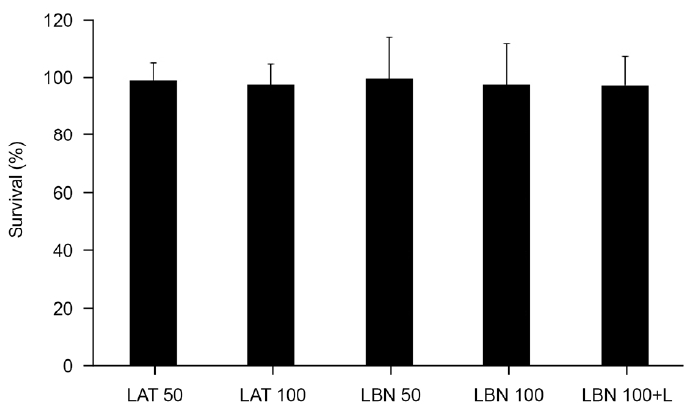
30ļČäĻ░ä ņĢĮļ¼╝ņŚÉ ļģĖņČ£ņŗ£Ēé© Ēøä ņäĖĒżņØś ņāØņĪ┤ņŚÉ ļ»Ėņ╣śļŖö ņśüĒ¢źņØä ņĖĪņĀĢĒĢśĻĖ░ ņ£äĒĢ┤ ņŗ£Ē¢ēĒĢ£ MTT assayņŚÉņä£ ņĢĮļ¼╝ņŚÉ ļģĖņČ£ņŗ£Ēéżņ¦Ć ņĢŖņØĆ ļīĆņĪ░ĻĄ░ņŚÉ ļ╣äĒĢ┤ LBNĻ│╝ LATļŖö Ļ░üĻ░ü ņäĖĒżņØś ņāØņĪ┤ņŚÉ ņ£ĀņØśĒĢ£ ņśüĒ¢źņØä ļ»Ėņ╣śņ¦Ć ņĢŖņĢśļŗż(all p>0.005) (Fig. 1). ļö░ļØ╝ņä£ ņĢäļלņØś Ēł¼Ļ│╝ļÅä ņŗżĒŚśņØś Ļ▓░Ļ│╝ļŖö ņäĖĒż ņłśņØś ļ│ĆĒÖöņŚÉ ņØśĒĢ£ Ļ▓āņØ┤ ņĢäļŗśņØä ņĢī ņłś ņ׳ņŚłļŗż.
LBNņØ┤ ņä¼ņ£ĀņŻ╝ņäĖĒżņØś NO ņāØņä▒ņŚÉ ļ»Ėņ╣śļŖö ņśüĒ¢ź
ņĢĮļ¼╝ņŚÉ ļģĖņČ£ņŗ£Ēéżņ¦Ć ņĢŖņØĆ ļīĆņĪ░ĻĄ░ņŚÉ ļ╣äĒĢśņŚ¼ 100 ╬╝M LBNņØĆ ļ░░ņ¦ĆņŚÉņä£ņØś nitrite ļåŹļÅäļź╝ ņ£ĀņØśĒĢśĻ▓ī ņ”ØĻ░Ćņŗ£ņ╝░ļŗż(p=0.029) (Fig. 2). 100 ╬╝M LBNņŚÉ L-NAMEļź╝ ĒĢ©Ļ╗ś ņ▓śļ”¼ĒĢ£ Ļ▓ĮņÜ░ LBN ļŗ©ļÅģņ£╝ļĪ£ ņ▓śļ”¼ĒĢ£ Ļ▓ĮņÜ░ņŚÉ ļ╣äĒĢ┤ nitriteņØś ļåŹļÅäĻ░Ć ņ£ĀņØśĒĢśĻ▓ī Ļ░ÉņåīĒĢśņśĆļŗż(p=0.041). LBNĻ│╝ LATļź╝ 50 ╬╝Mņö® ņ▓śļ”¼ĒĢ£ Ļ▓ĮņÜ░ļź╝ ļ╣äĻĄÉĒĢ┤ļ│┤ļ®┤ NO ņāØņä▒ņŚÉņä£ ņ£ĀņØśĒĢ£ ņ░©ņØ┤ļź╝ ļ│┤ņØ┤ņ¦Ć ņĢŖņĢśņ£╝ļéś(p=0.191), LBNĻ│╝ LATļź╝ 100 ╬╝Mņö® ņ▓śļ”¼ĒĢ£ Ļ▓ĮņÜ░ļź╝ ļ╣äĻĄÉĒĢ┤ļ│┤ļ®┤ LBNņØ┤ LATņŚÉ ļ╣äĒĢ┤ ņ£ĀņØśĒĢśĻ▓ī nitriteņØś ļåŹļÅäļź╝ ņ”ØĻ░Ćņŗ£ņ╝░ļŗż(p=0.040).
LBNņØ┤ ņä¼ņ£ĀņŻ╝ļŗ©ņĖĄņäĖĒżņĖĄņØś Ēł¼Ļ│╝ļÅäņŚÉ ļ»Ėņ╣śļŖö ņśüĒ¢ź
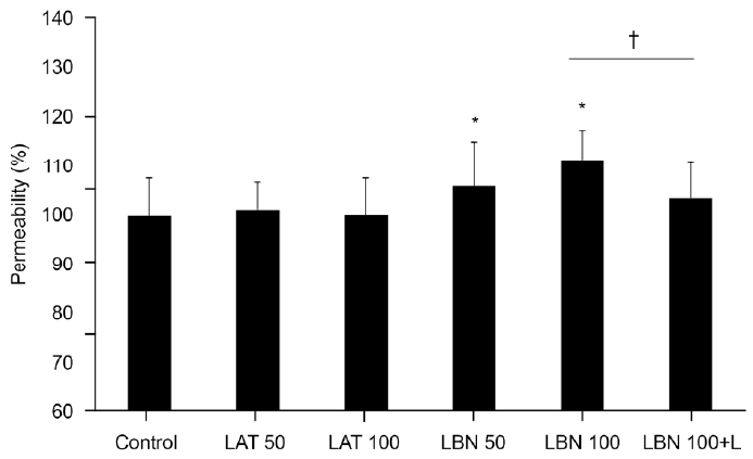
ņĢĮļ¼╝ņŚÉ ļģĖņČ£ņŗ£Ēéżņ¦Ć ņĢŖņØĆ ļīĆņĪ░ĻĄ░ņŚÉ ļ╣äĒĢśņŚ¼ 50, 100 ╬╝M LBNņØĆ carboxyfluoresceinņØś ņä¼ņ£ĀņŻ╝ļŗ©ņĖĄņäĖĒżņĖĄņØś Ēł¼Ļ│╝ļÅäļź╝ 106.1%, 111.4%ļĪ£ Ļ░üĻ░ü ņ£ĀņØśĒĢśĻ▓ī ņ”ØĻ░Ćņŗ£ņ╝░ņ£╝ļéś(p=0.010, 0.001) LATļŖö ņ£ĀņØśĒĢ£ ņśüĒ¢źņØä ļ»Ėņ╣śņ¦Ć ņĢŖņĢśļŗż(Fig. 3). 100 ╬╝M LBNņŚÉ L-NAMEļź╝ ĒĢ©Ļ╗ś ņ▓śļ”¼ĒĢ£ Ļ▓ĮņÜ░ 100 ╬╝M LBN ļŗ©ļÅģņ£╝ļĪ£ ņ▓śļ”¼ĒĢ£ Ļ▓ĮņÜ░ņŚÉ ļ╣äĒĢ┤ Ēł¼Ļ│╝ļÅäĻ░Ć 7.8% Ļ░ÉņåīĒĢśņśĆļŗż(p=0.001). LBNĻ│╝ LATļź╝ ņ▓śļ”¼ĒĢ£ Ļ▓ĮņÜ░ Ēł¼Ļ│╝ļÅäļź╝ ļ╣äĻĄÉĒĢ┤ļ│┤ļ®┤ 50, 100 ╬╝M ļåŹļÅä ļ¬©ļæÉņŚÉņä£ LATņŚÉ ļ╣äĒĢ┤ LBNņØ┤ ņ£ĀņØśĒĢśĻ▓ī Ēł¼Ļ│╝ļÅäļź╝ ņ”ØĻ░Ćņŗ£ņ╝░ļŗż(p=0.018, 0.001).
LBNņØ┤ ņä¼ņ£ĀņŻ╝ļŗ©ņĖĄņäĖĒżņĖĄņØś ņĀĆĒĢŁļÅäņŚÉ ļ»Ėņ╣śļŖö ņśüĒ¢ź
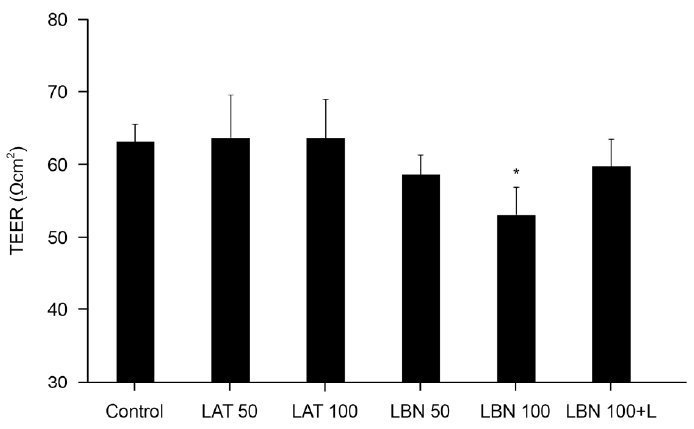
ņĢĮļ¼╝ņŚÉ ļģĖņČ£ņŗ£Ēéżņ¦Ć ņĢŖņØĆ ļīĆņĪ░ĻĄ░ņØś ņĀĆĒĢŁļÅäļŖö 63.02 Ōä” cm2ņśĆņ£╝ļ®░ ņØ┤ņŚÉ ļ╣äĒĢśņŚ¼ 100 ╬╝M LBNņØä ņ▓śļ”¼ĒĢ£ Ļ▓ĮņÜ░ņØś ņĀĆĒĢŁļÅäļŖö 53.01 Ōä” cm2ļĪ£ 15.89% Ļ░ÉņåīĒĢśņśĆļŗż(p=0.047) (Fig. 4). 50 ╬╝M LBNņØä ņ▓śļ”¼ĒĢ£ Ļ▓ĮņÜ░ ņĀĆĒĢŁļÅäļŖö 58.48 Ōä” cm2ļĪ£ 7.21% Ļ░ÉņåīĒĢśņśĆņ£╝ļéś ĒåĄĻ│äņĀüņ£╝ļĪ£ ņ£ĀņØśĒĢśņ¦ĆļŖö ņĢŖņĢśņ£╝ļ®░(p=0.235), 50 ļśÉļŖö 100 ╬╝M LATļŖö ļīĆņĪ░ĻĄ░ņŚÉ ļ╣äĒĢśņŚ¼ ņĀĆĒĢŁļÅäņØś ņ£ĀņØśĒĢ£ ņ░©ņØ┤ļź╝ ļ│┤ņØ┤ņ¦Ć ņĢŖņĢśļŗż.
100 ╬╝M LBNņŚÉ L-NAMEļź╝ ĒĢ©Ļ╗ś ņ▓śļ”¼ĒĢ£ Ļ▓ĮņÜ░ 100 ╬╝M LBN ļŗ©ļÅģņ£╝ļĪ£ ņ▓śļ”¼ĒĢ£ Ļ▓ĮņÜ░ņŚÉ ļ╣äĒĢ┤ ņĀĆĒĢŁļÅäļŖö 10.58% Ļ░ÉņåīĒĢśņśĆņ£╝ļéś ĒåĄĻ│äņĀüņ£╝ļĪ£ ņ£ĀņØśĒĢśņ¦Ć ņĢŖņĢśņ£╝ļ®░(p=0.162), LBNĻ│╝ LATļź╝ ņ▓śļ”¼ĒĢ£ Ļ▓ĮņÜ░ņØś Ēł¼Ļ│╝ļÅäļź╝ Ļ░üĻ░ü ļ╣äĻĄÉĒĢ┤ļ│┤ļ®┤ 50, 100 ╬╝M ļåŹļÅä ļ¬©ļæÉņŚÉņä£ LATņŚÉ ļ╣äĒĢ┤ LBNņØ┤ ļåŹļÅäĻ░Ć ņ”ØĻ░ĆĒĢĀņłśļĪØ ņĀĆĒĢŁļÅäļź╝ Ļ░Éņåīņŗ£ĒéżļŖö Ļ▓ĮĒ¢źņØä ļéśĒāĆļé┤ņŚłņ£╝ļéś ĒåĄĻ│äņĀüņ£╝ļĪ£ ņ£ĀņØśĒĢśņ¦ĆļŖö ņĢŖņĢśļŗż(p=0.444, 0.073).
Ļ│Ā ņ░░
ĻĖ░ņĪ┤ņØś ļÅÖļ¼╝ņŗżĒŚśņŚÉņä£ ĒöäļĪ£ņŖżĒāĆĻĖĆļ×ĆļöśņŚÉ NOļź╝ Ļ▓░ĒĢ®ņŗ£Ēé© ņĢĮņĀ£ņØś ņĢłņĢĢĒĢśĻ░Ģ ĒÜ©Ļ│╝Ļ░Ć ļ│┤Ļ│ĀļÉ£ ņØ┤Ēøä11,35 ņĄ£ĻĘ╝ Ļ░£ļ░£ļÉ£ LBNņØĆ ĻĖ░ņĪ┤ņØś ļģ╣ļé┤ņן ņĢłņĢĢĒĢśĻ░ĢņĀ£ļōżņŚÉ ļ╣äĒĢ┤ ņÜ░ņłśĒĢ£ ņĢłņĢĢĒĢśĻ░Ģ ĒÜ©Ļ│╝ļź╝ ļéśĒāĆļé┤ņŚłļŗżļŖö ņŚ¼ļ¤¼ ņ×äņāü ņŚ░ĻĄ¼ Ļ▓░Ļ│╝ļōżņØ┤ ļ│┤Ļ│ĀļÉśņ¢┤ ņÖöļŗż.12-20 ņØ┤ļ¤¼ĒĢ£ LBNņØś ņÜ░ņłśĒĢ£ ĒÜ©Ļ│╝ļŖö ļÅÖļ¼╝ņŗżĒŚśņØä ĒåĄĒĢ┤ņä£ļŖö LATņŚÉ ņØśĒĢ£ ĒżļÅäļ¦ēĻ│Ąļ¦ēņ£ĀņČ£ņØä ņ┤ēņ¦äĒĢśļŖö ņ×æņÜ®Ļ│╝ ņä¼ņ£ĀņŻ╝ļź╝ ĒåĄĒĢ£ ļ░®ņłśņ£ĀņČ£ņØä ņ”ØĻ░Ćņŗ£ĒéżļŖö NOņØś ņØ┤ņżæ ņ×æņÜ®ņŚÉ ņØśĒĢ£ Ļ▓āņ£╝ļĪ£ ņŚ¼Ļ▓©ņĀĖ ņÖöņ¦Ćļ¦ī,11 LBNņØ┤ ņØĖņ▓┤ ļé┤ņŚÉņä£ ņä¼ņ£ĀņŻ╝ņØś ļ░®ņłśņ£ĀņČ£ņŚÉ ļ»Ėņ╣śļŖö ņśüĒ¢źņØĆ ņä¼ņ£ĀņŻ╝ņäĖĒżļź╝ ņØ┤ņÜ®ĒĢ£ ņŗżĒŚśņŗż ļé┤ ņŚ░ĻĄ¼ņŚÉņä£ļŖö ņĢäņ¦ü ņ×ÉņäĖĒ׳ ļ░ØĒśĆņ¦Ćņ¦Ć ņĢŖņĢśļŗż. ņØ┤ņŚÉ ļö░ļØ╝ ļ│Ė ņŚ░ĻĄ¼ņŚÉņä£ļŖö ļ░░ņ¢æĒĢ£ ņØĖņ▓┤ ņä¼ņ£ĀņŻ╝ņäĖĒżļź╝ ņØ┤ņÜ®ĒĢśņŚ¼ ņŗżĒŚśņŗż ļé┤ņŚÉņä£ LBNņØ┤ ņä¼ņ£ĀņŻ╝ļź╝ ĒåĄĒĢ£ ļ░®ņłśņ£ĀņČ£ņŚÉ ļ»Ėņ╣śļŖö ņśüĒ¢źņØä LATņÖĆ ļ╣äĻĄÉĒĢśņŚ¼ ņĢīņĢäļ│┤ņĢśļŗż.
ļ│Ė ņŚ░ĻĄ¼ņØś Ļ▓░Ļ│╝ņŚÉņä£ LBNņØĆ ņä¼ņ£ĀņŻ╝ļŗ©ņĖĄņäĖĒżņĖĄņØś Ēł¼Ļ│╝ļÅäļź╝ ļåŹļÅäņŚÉ ļ╣äļĪĆĒĢśņŚ¼ ņ”ØĻ░Ćņŗ£ņ╝░ņ£╝ļ®░ ņĀĆĒĢŁļÅäļź╝ Ļ░Éņåīņŗ£ņ╝░ņ£╝ļ»ĆļĪ£ LBNņØ┤ ņä¼ņ£ĀņŻ╝ļź╝ ĒåĄĒĢ£ ļ░®ņłśņ£ĀņČ£ņØä ņ”ØĻ░Ćņŗ£ĒéżļŖö ņ×æņÜ®ņØä ļéśĒāĆļāäņØä ĒÖĢņØĖĒĢĀ ņłś ņ׳ņŚłļŗż. ņØ┤ņŚÉ ļ╣äĒĢ┤ LATļŖö Ēł¼Ļ│╝ļÅäņŚÉ ņ£ĀņØśĒĢ£ ņśüĒ¢źņØä ļ»Ėņ╣śņ¦Ć ņĢŖņĢśļŖöļŹ░ ĻĖ░ņĪ┤ņØś ļ¬ćļ¬ć ņŚ░ĻĄ¼ņŚÉņä£ ļØ╝ĒāĆļģĖĒöäļĪ£ņŖżĒŖĖĻ░Ć ņä¼ņ£ĀņŻ╝ņŚÉņä£ ņäĖĒżņÖĖĻĖ░ņ¦łņØä Ļ░Éņåīņŗ£ĒéżĻ▒░ļéś ĻĖ░ņ¦łĻĖłņåŹļŗ©ļ░▒ņ¦łļČäĒĢ┤ĒÜ©ņåīļź╝ ņ”ØĻ░Ćņŗ£ņ╝£ ņä¼ņ£ĀņŻ╝ļź╝ ĒåĄĒĢ£ ļ░®ņłśņ£ĀņČ£ņØä ņ┤ēņ¦äņŗ£Ēé©ļŗżļŖö ļ│┤Ļ│ĀļÅä ņ׳ņŚłņ£╝ļéś4,36 ņØ┤ņŚÉ ļīĆĒĢ┤ņä£ļŖö ņĢäņ¦ü ļģ╝ļ×ĆņØś ņŚ¼ņ¦ĆĻ░Ć ļ¦ÄļŗżĻ│Ā ļ│╝ ņłś ņ׳ņ£╝ļ®░ ļ│Ė ņŚ░ĻĄ¼ņØś Ļ▓░Ļ│╝ņŚÉ ļö░ļź┤ļ®┤ LATļŖö ņä¼ņ£ĀņŻ╝ļź╝ ĒåĄĒĢ£ ļ░®ņłśņ£ĀņČ£ņŚÉļŖö ņ£ĀņØśĒĢ£ ņśüĒ¢źņØä ļ»Ėņ╣śņ¦Ć ņĢŖņĢśņ£╝ļ»ĆļĪ£ LATĻ░Ć ņä¼ņ£ĀņŻ╝ļź╝ ĒåĄĒĢ£ ļ░®ņłśņ£ĀņČ£ņŚÉ ļ»Ėņ╣śļŖö ņśüĒ¢źņØĆ ņĀüņØä Ļ▓āņ£╝ļĪ£ ņāØĻ░üļÉ£ļŗż. ļö░ļØ╝ņä£ LBNņØĆ LATņŚÉ ļ╣äĒĢ┤ ņä¼ņ£ĀņŻ╝ļź╝ ĒåĄĒĢ£ ļ░®ņłśņ£ĀņČ£ņØś ņ”ØĻ░Ćļź╝ ņČöĻ░ĆņĀüņ£╝ļĪ£ ļéśĒāĆļé╝ ņłś ņ׳ņ£╝ļ»ĆļĪ£ ņĪ░ĻĖł ļŹö ņÜ░ņłśĒĢ£ ņĢłņĢĢĒĢśĻ░Ģ ĒÜ©Ļ│╝ļź╝ ļéśĒāĆļé╝ Ļ▓āņ£╝ļĪ£ ņŚ¼Ļ▓©ņ¦äļŗż.
ņØ┤ļ¤¼ĒĢ£ LBNņØś ņČöĻ░ĆņĀüņØĖ ņĢłņĢĢĒĢśĻ░Ģ ĒÜ©Ļ│╝ļŖö ņä¼ņ£ĀņŻ╝ņŚÉ ņ×æņÜ®ĒĢśļŖö NOņŚÉ ņØśĒĢ£ Ļ▓āņ£╝ļĪ£ ņŚ¼Ļ▓©ņ¦Ćļ®░37 ļ│Ė ņŚ░ĻĄ¼ņä£ļÅä LBNņØ┤ NOņØś ņāØņä▒ņØä ņ”ØĻ░Ćņŗ£ĒéżļŖö Ļ▓āņ£╝ļĪ£ ļéśĒāĆļé¼ļŗż. NOļŖö cGMPļź╝ ĒåĄĒĢśņŚ¼ ņ×æņÜ®ņØä ļéśĒāĆļé┤ļŖöļŹ░21-23 ļ│Ė ņŚ░ĻĄ¼ņŚÉņä£ cGMP ņĀ£ĒĢ┤ņĀ£ļĪ£ ņ×æņÜ®ĒĢśļŖö L-NAMEļź╝38,39 ĒĢ©Ļ╗ś ļģĖņČ£ņŗ£Ēé© Ļ▓░Ļ│╝ LBNņŚÉ ņØśĒĢ£ ņä¼ņ£ĀņŻ╝ļŗ©ņĖĄņäĖĒżņĖĄņØś Ēł¼Ļ│╝ļÅä ņ”ØĻ░ĆņÖĆ ņĀĆĒĢŁļÅä Ļ░ÉņåīĻ░Ć ņāüņćäļÉśņŚłļŗż. ļö░ļØ╝ņä£ LBNņØĆ ņä¼ņ£ĀņŻ╝ņŚÉņä£ NOņØś ņāØņä▒ņØä ņ”ØĻ░Ćņŗ£ņ╝£ cGMPļź╝ ĒåĄĒĢ┤ ņä¼ņ£ĀņŻ╝ļź╝ ņØ┤ņÖäņŗ£ņ╝£ ļ░®ņłśņ£ĀņČ£ņØä ņ┤ēņ¦äņŗ£ĒéżļŖö ņ×æņÜ®ņØä ļéśĒāĆļéĖļŗżĻ│Ā ļ│╝ ņłś ņ׳Ļ▓Āļŗż.
ļ╣äļĪØ ļ│Ė ņŚ░ĻĄ¼Ļ░Ć ņŗżĒŚśņŗż ļé┤ņŚÉņä£ ņä¼ņ£ĀņŻ╝ļŗ©ņĖĄņäĖĒżņĖĄņØä ņØ┤ņÜ®ĒĢ£ Ēł¼Ļ│╝ļÅäļź╝ ļ╣äĻĄÉ ņŚ░ĻĄ¼ĒĢ£ Ļ▓░Ļ│╝ņØ┤ĻĖ░ļŖö ĒĢśņ¦Ćļ¦ī ņä¼ņ£ĀņŻ╝ņäĖĒżļź╝ ņØ┤ņÜ®ĒĢ£ ņØ┤ņĀäņØś ņŗżĒŚśņŗż ļé┤ ņŚ░ĻĄ¼ņŚÉņä£ LBNņØ┤ LATņŚÉ ļ╣äĒĢ┤ ņ£ĀņØśĒĢśĻ▓ī ņä¼ņ£ĀņŻ╝ļź╝ ņØ┤ņÖäņŗ£ņ╝░ļŗżļŖö Ļ▓░Ļ│╝ļź╝24 ĒĢ©Ļ╗ś Ļ│ĀļĀżĒĢ┤ļ│┤ļ®┤ LBNņØĆ ĒżļÅäļ¦ēĻ│Ąļ¦ēņ£ĀņČ£Ļ│╝ ņä¼ņ£ĀņŻ╝ņ£ĀņČ£ņØä ĒĢ©Ļ╗ś ņ┤ēņ¦äĒĢśļŖö ņØ┤ņżæ ņ×æņÜ®ņØä ļéśĒāĆļé┤ļ®░, ņØ┤ņŚÉ ļö░ļØ╝ ĻĖ░ņĪ┤ņØś ļØ╝ĒāĆļģĖĒöäļĪ£ņŖżĒŖĖ ņĀ£ņ×¼ļ│┤ļŗż ņ×äņāüņĀüņ£╝ļĪ£ ņÜ░ņłśĒĢ£ ņĢłņĢĢĒĢśĻ░Ģ ĒÜ©Ļ│╝ļź╝ ļéśĒāĆļéĖļŗżļŖö ņØ┤ņĀä ņ×äņāü ņŚ░ĻĄ¼ļōżņØś Ļ▓░Ļ│╝ļōżņØä ļÆĘļ░øņ╣©ĒĢ£ļŗż.
ļśÉĒĢ£ NOļŖö ņĢłņĢĢĒĢśĻ░Ģ ĒÜ©Ļ│╝ļ┐Éļ¦ī ņĢäļŗłļØ╝ ņé░ĒÖöņŖżĒŖĖļĀłņŖżļź╝ Ļ░Éņåīņŗ£Ēéżļ®░ ņŗ£ņŗĀĻ▓ĮĒśłļźśļź╝ ņ”ØĻ░Ćņŗ£ņ╝£ ņŗĀĻ▓Įļ│┤ĒśĖ ĒÜ©Ļ│╝ļź╝ ļéśĒāĆļéĖļŗżĻ│Ā ņĢīļĀżņĀĖ ņ׳ļŖöļŹ░,40 ņØ┤ņŚÉ ļīĆĒĢ£ LBNņØś ņ×æņÜ®ņŚÉ Ļ┤ĆĒĢ┤ņä£ļŖö Ē¢źĒøä ļŹö ņ×ÉņäĖĒĢ£ ņŚ░ĻĄ¼Ļ░Ć ĒĢäņÜöĒĢĀ Ļ▓āņØ┤ļŗż.
Ļ▓░ļĪĀņĀüņ£╝ļĪ£ ņä¼ņ£ĀņŻ╝ņäĖĒżņŚÉņä£ LBNņØĆ ļØ╝ĒāĆļģĖĒöäļĪ£ņŖżĒŖĖņŚÉ ņØśĒĢ£ ĒżļÅäļ¦ēĻ│Ąļ¦ēņ£ĀņČ£ ņ”ØĻ░ĆņÖĆ ļŹöļČłņ¢┤ NOņŚÉ ņØśĒĢ£ ņä¼ņ£ĀņŻ╝ņ£ĀņČ£ņØä ĒĢ©Ļ╗ś ņ”ØĻ░Ćņŗ£Ēé┤ņ£╝ļĪ£ņŹ© ĻĖ░ņĪ┤ņØś ļØ╝ĒāĆļģĖĒöäļĪ£ņŖżĒŖĖ ņĀ£ņĀ£ļ│┤ļŗż ņĪ░ĻĖł ļŹö ņÜ░ņłśĒĢ£ ņĢłņĢĢĒĢśĻ░Ģ ņ×æņÜ®ņØä ļéśĒāĆļé╝ ņłś ņ׳ņØä Ļ▓āņ£╝ļĪ£ ņāØĻ░üļÉ£ļŗż.

 PDF Links
 PubReader
 ePub Link
 Full text via DOI
 Download Citation
 Print
Print


