눈꺼풀의 발적성 부종을 주소로 내원한 소아 피부근육염 1예
Juvenile Dermatomyositis Presenting with Erythematous Swelling of the Upper Eyelid
Article information
Abstract
목적
눈꺼풀부종으로 내원한 환아에서 소아 피부근육염으로 진단된 1예를 보고하고자 한다.
증례요약
11세 여자 환아가 3주 전부터 시작된 양측 위눈꺼풀부종으로 내원하였다. 양안 통증, 충혈은 없었으며 양 뺨의 발진이 있었다. 눈꺼풀염 의심하 항염증 치료 중 위눈꺼풀테 주변으로 적자색의 헬리오트로프 발진 양상이 확인되었다. 발열, 관절통, 구강내 궤양 및 귀 뒤의 인설성 홍반과 손가락 관절과 팔꿈치에 인설성 홍반성 구진도 동반되었다. 혈청검사상 aspartate transaminase 184 IU/L, alanine transaminase 352 IU/L, alkaline phosphatase 266 IU/L로 상승하였다. 조직검사상 눈꺼풀 병변은 피부근육염, 손가락 관절 병변은 고트론 구진으로 확인되었다. European League Against Rheumatism/American College of Rheumatology 진단 기준에 따라 소아 피부근육염으로 진단되어 스테로이드 치료하던 중 합병증으로 발생한 간질성 폐렴이 급격히 진행하여 진단 3개월에 사망하였다.
결론
눈꺼풀부종 환자에서 헬리오트로프 발진을 확인하여 소아 피부근육염의 조기 진단 가능성을 높일 수 있다.
Trans Abstract
Purpose
To report a case of juvenile dermatomyositis that presented with erythematous swelling on the upper eyelid as the first clinical sign.
Case summary
An 11-year-old female patient presented with erythematous swelling of both upper eye lids that persisted for 3 weeks prior to examination. She also had an erythematous rash over both cheeks but no pain or hyperemia in either eye. During anti-inflammatory eye drop treatment under suspicion of blepharitis, a reddish-purple heliotrope rash appeared around the eyelid margin. She also developed fever, arthralgia, multiple oral ulcers, an erythematous scaly rash behind the ear, and erythematous firm papules on the metacarpal (MCP) joint and around the elbow. On evaluation, aspartate aminotransferase was elevated to 184 IU/L, alanine aminotransferase to 352 IU/L, and alkaline phosphatase to 266 IU/L. We performed a skin biopsy. Based on histopathological examination, the eyelid lesion was diagnosed as dermatomyositis and the MCP joint lesion as Gottron’s papule. From the above findings, we diagnosed the patient with juvenile dermatomyositis based on the European League Against Rheumatism/American College of Rheumatology classification. The patient was treated with prednisolone. Three months after diagnosis, she died from rapidly progressive interstitial pneumonia, a complication of juvenile dermatomyositis.
Conclusions
Heliotrope rash should be considered in the differential diagnosis of patients presenting with eyelid swelling, for early diagnosis and better prognosis of juvenile dermatomyositis.
소아 피부근육염(Juvenile dermatomyositis)은 전신혈관병증을 특징으로 하는 자가면역 질환이다[1]. 만성화될수록 피부 및 근육의 석회화로 기능이 제한되거나 장기의 축적성 손상이 올 확률이 높으나, 조기에 치료를 시작할수록 만성화될 확률이 낮으며 좋은 예후를 갖는 것으로 알려져 있다[2-4].
소아 피부근육염의 가장 대표적인 증상은 특징적인 피부발진(Heliotrope rash, Gottron’s papule, Gottron’s sign)과 근위부 근력 약화가 있고, 그 외에도 소화기, 폐, 심장, 관절, 시각, 신경에 증상을 일으킬 수 있다[5]. 눈꺼풀의 발적성부종은 소아 피부근육염 환자의 83%에서 나타나는 것으로 알려져 있으나[5], 국내에서 소아 환자가 안병변을 첫 증상으로 내원하여 소아 피부근육염을 진단하여 보고한 사례는 없었다. 이에 저자들은 양측 위눈꺼풀의 부종을 첫 주소로 내원한 환자에서, 경과 관찰 도중 헬리오트로프 발진이 확인되어 소아 피부근육염이 최종 진단된 1례를 경험하였기에 이를 보고하는 바이다.
증례보고
기저질환이 없는 11세 여자 환아가 3주 전부터 시작된 양측 위눈꺼풀의 부종이 보존적 치료에 호전이 없어 내원하였다. 특이 과거력은 없었다. 내원시 양측 뺨 전체에 발진이 동반되었고 양안 통증, 충혈 및 삼출물은 없었다. 양안 시력 1.0이었고, 안압은 우안 12 mmHg, 좌안 13 mmHg였으며, 전안부검사상 특이 소견은 보이지 않았다.
눈꺼풀염 진단하에 안연고(Dexamethasone, Neomycin Sulfate, Polymyxin B Sulfate)를 처방하여 일주일 후 부종은 일부 호전되었으나, 구강 내에는 여러 개의 궤양이 발생하여(Fig. 1) 혈액검사를 시행하였다. 혈액검사상 aspartate transaminase (AST) 184 IU/L, alanine transaminase (ALT) 352 IU/L, alkaline phosphatase (ALP) 266 IU/L로 상승 소견을 보였으며, antinuclear antibody 음성 소견, erythrocyte sedimentation rate 14 mm/hr, C-reactive protein 0.19 mg/dL로 정상 소견을 보였다.
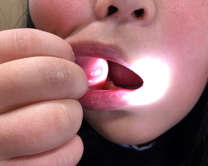
Clinical photograph of oral ulcer of the 11-year-old patient. One week after treatment of anti-inflammatory eye ointment (Dexamethasone, Neomycin Sulfate, Polymyxin B Sulfate), multiple oral ulcer was observed.
내원 9일 후, 귀 뒤편 골 돌출부에 인설성 홍반(Fig. 2A)이 관찰되었고 손가락의 중수지 관절과 팔꿈치에 인설성 홍반이 융기된 양상의 병변이 생겼다(Fig. 2B). 위눈꺼풀부종은 일부 호전되면서 적자색의 헬리오트로프 발진에 합당한 소견이 보였다(Fig. 3). 피부과 협진하 시행한 조직검사에서 눈꺼풀의 병변은 피부근육염(dermatomyositis) 소견이 나왔고, 중수지 관절의 병변은 고트론 구진(Gottron’s papule)에 합당한 소견이 확인되었다(Fig. 4). 또한 환아는 추가로 손목, 발목, 손가락의 관절통을 호소하였고 37.7°C의 발열이 있었다. 이에 소아청소년과 협진하에 항생제와 프레드니솔론 복용 치료를 시작하였다.

Clinical photograph of skin rash over bony prominences. (A) Erythematous scaly rash appeared after one week of treatment, which localized in bony prominences around ear. (B) Erythematous, papulosquamous eruption appeared over the both elbow, which are called Gottron’s papules.

Clinical photograph of the face. After one week of treatment, reddish-purple swelling of both eyelid which seemed to be Heliotrope rash and erythematous facial rash appeared. The patient consented to the use of this photograph.

Histological sections of biopsy of skin, eyelid. Histological sections of skin biopsy show mild hyperkeratosis with epidermal atrophy, diffuse basement membrane thickening and basal vacuolar change (arrows). Minimal perivascular lymphocytic infiltration is limited to superficial dermis (hematoxylin and eosin staining, ×200).
내원 21일 뒤 추가로 시행한 혈액검사상 AST 303 IU/L, ALT 254 IU/L, lactate dehydrogenase (LDH) 584 U/L, creatine phosphokinase 428 U/L로 상승 소견을 보였다. 근전도검사(electromyography)상에서는 명확한 근병증 소견은 보이지 않았으나, 헬리오트로프 발진, 고트론 구진, AST, ALT 상승 소견으로 European League Against Rheumatism/American College of Rheumatology (EULAR/ACR) [6]의 소아 피부근육염 진단 기준을 만족하여 소아 피부근육염으로 진단하 프레드니솔론 치료를 유지하였다.
내원 2개월째 양팔과 다리의 힘 빠짐 증상과 38.3°C의 발열로 입원하여 정맥내 메틸프레드니솔론(methylprednisolone) 치료 후 퇴원하였다. 이후 경구 프레드니솔론, 아자티오프린(Azathioprine) 치료받던 중 숨차는 증상과 기침이 심해져 간질성 폐렴(interstitial lung disease) 진단하 입원하여 항생제 투여 치료받았으나, 체외막산소공급(extracorporeal membrane oxygenation) 치료 중 사망하였다.
고 찰
소아 피부근육염은 주로 피부와 근골격계에 염증을 일으켜 근위부 근력약화와 발진을 일으키는, 원인이 명확히 밝혀지지 않은 전신 자가면역질환이다[1]. 소아 피부근육염의 발병률은 한국의 발병률은 잘 알려져 있지 않으나 미국은 매년 백만 명당 2.5-4.1명으로 알려져 있다[7].
다양한 임상증상이 나타나는데 특징적으로는 홍반성 구진 인설성 발진(고트론 발진 및 징후[Gottron’s rash], 91%), 눈꺼풀의 적자색 발진성 부종(헬리오트로프 발진[Heliotrope rash], 83%), 얼굴 발진(malar/facial rash, 42%)이 나타나며, 그 외 손톱 모세혈관 변화(80%), 근육/관절통(25%), 발성/연하장애(24%), 식욕부진(18%), 발열(16%) 등이 나타난다[5]. 2005년 Akikusa et al [8]에 따르면 그중 눈의 증상으로는 눈꺼풀피부의 변화가 가장 흔하며, 그 외 스테로이드 치료로 인한 백내장과 드물게 망막병증 또한 나타날 수 있다. 눈꺼풀 피부에 나타나는 전형적인 헬리오트로프 발진은 눈꺼풀의 넓게 퍼진 적자색의 홍반으로 부종을 동반하지 않을 수 있고 뺨, 광대뼈 부위(malar region)까지 이어지기도 하며 활동성으로 염증성 혈관병증이 일어나고 있다는 것을 의미한다.
본 증례의 환자는 눈꺼풀의 부종을 첫 증상으로 내원하여 진료 시 뺨/광대뼈 부위 발진이 동반되는 것을 발견한 후, 경과 관찰 중 헬리오트로프 발진이 확인되었고 발열, 관절통, 구강 궤양, 귀 뒤 피부 발진 및 중수지관절과 팔꿈치의 고트론 구진이 추가로 나타난 경우이다. 소아 피부근육염 환자의 혈액검사에서는 근 손상으로 인해 Creatinine kinase (CK), LDH, ALT, AST, ALP, Aldolase에서 상승 소견이 첫 증상 발생 2.53-4.65개월 내에 나타나는 것으로 알려져 있으며[9] 본 증례의 환자에서도 AST, ALT, LDH, CK, ALP, Aldolase의 상승 소견을 보였다.
2017년 EULAR/ACR에서 제안한 진단 기준에 따르면 근육 생검을 하지 않은 경우: 1) 첫 증상이 나타난 나이가 18세 이하인 경우(1.3점), 40세 이상인 경우(2.1점), 2) 근력 약화 양상이 상지 근위부 대칭성인 경우(0.7점), 하지 근위부 대칭성인 경우(0.8점), 목 굴곡근이 신전근보다 약한 경우(1.9점), 하지 근위부 근육이 원위부 근육보다 약한 경우(0.9점), 3) 적자색 발진이 눈꺼풀이나 눈 주변에 나타난 경우(헬리오트로프 발진[Heliotrope rash], 3.1점), 손가락, 팔꿈치, 무릎, 발목, 발가락 등에 홍반성 구진 인설성 발진(고트론 구진[Gottron papules], 2.1점) 홍반성 구진이 아닌 인설성 발진(고트론 징후[Gottron sign], 3.3점), 4) 연하 곤란 또는 식도기능부전(0.7점), 5) 혈액검사상 Anti-Jo-1 양성(3.9점), CK 또는 LDH 또는 AST 또는 ALT 상승(1.3점) 각각의 점수의 합이 7.5점 이상인 경우 명확한 특발성 염증성 근병증(idiopathic inflammatory myopathies), 5.5점에서 7.5점 사이인 경우 가능성 높은 특발성 염증성 근병증(probable idiopathic inflammatory myopathies), 5.3점에서 5.4점 사이인 경우 가능성 있는 특발성 염증성 근병증(possible idiopathic inflammatory myopathies)으로 정의하였으며, 그중 세 가지 특징적인 피부 증상인 헬리오트로프 발진, 고트론 구진, 고트론 징후가 있는 경우 소아 피부근육염으로 진단된다[6]. 근생검과 근전도검사는 침습적이기 때문에 소아 피부근육염의 특징적인 임상증상이 있는 경우에서는 잘 사용되지 않으며, 최근에는 임상증상으로 주로 진단하는 추세이다[10]. 본 증례의 환자 또한 첫 증상이 나타난 나이가 18세 이하(1.3점), 헬리오트로프 발진(3.1점), 고트론 구진(2.1점), AST와 ALT 상승(1.3점)을 만족하여 7.8점으로 소아 피부근육염으로 진단되었다. 또한 이후 시행한 조직검사상에서도 눈꺼풀 병변에서는 피부근육염(dermatomyositis) 소견과 손가락 중수지절관절의 병변에서는 고트론 구진 소견으로, 소아 피부근육염 진단에 합당한 소견을 받았다.
소아 피부근육염의 일차 치료는 경구 고용량 스테로이드로 알려져 있으며, 스테로이드의 사용량을 줄이기 위해 메토트렉세이트(methotrexate)와 사이클로스포린(cyclosporine) 등이 같이 사용될 수 있다[11]. 소아 피부근육염에서 조기에 시작된 올바른 치료가 질병의 경과를 재발 없이 일반적인 치료법에 반응하도록 하여, 만성화보다 좋은 예후를 갖게 하는 것으로 알려져 있다[2,3].
Pachman et al [9]은 79명의 소아 피부근육염 환아들을 대상으로 한 조사에서 처음 증상이 나타날 때부터 진단까지의 기간이 평균 3개월이며, 가장 기간이 긴 경우가 20개월이라 보고하였다. 이는 발열, 근육 약화 같은 초기 증상들이 비특이적이어서 지나치기 쉽고, 특징적인 피부 발진 소견과 근육 약화가 나타나더라도 단순 안검염이나 피부 질환으로 오인해 진단할 수 있기 때문으로 생각된다. 본 증례의 환자는 증상이 시작된 지 약 3주 후 내원하여, 증상 발현 약 6주만에 비교적 조기 진단되어 경구 프레드니솔론 치료를 시작할 수 있었다.
소아 피부근육염은 세 가지 패턴의 경과를 보인다. 1/3의 환자들이 2-3년간 지속되는 단주기성 경과를 가지며 적절한 치료 시 재발하지 않는다. 약 3%의 환자들은 관해와 재발이 반복되는 다주기성 경과를 가지며 2/3의 환자들은 만성 지속성의 경과를 밟으며 영구적인 합병증을 갖기도 한다[12]. 만성적이고 지속적인 경과는 좋지 않은 예후의 가장 큰 예측인자로 알려져 있다[3]. 소아 피부근육염은 다발성 장기 손상을 일으키는 자가면역질환으로, 피부 및 근골격계 다음으로 폐가 가장 많이 손상받는 장기이다. 75%의 소아 피부근육염 환아에서 호흡기계 손상이 일어나며, 이는 호흡기 근육약화, 면역 억제 치료, 간질성 폐렴 등으로 인해 일어난다. 간질성 폐렴은 8-19%의 소아 피부근육염 환자에서 일어나며, 소아 피부근육염으로 인한 사망의 주된 원인으로 알려져 있다. 그리고 심장 손상으로 인한 임상적 이상 증상을 보이지 않는 환자에서도 심전도와 심초음파상에서는 이상 소견을 보일 수 있다. 또한 드물지만 소화기계, 신경계 이상 또한 혈관병증으로 인해 생길 수 있다[13]. 스테로이드 치료가 시작되기 전 소아 피부근육염의 사망률은 30% 이상이었으나 스테로이드 치료의 도입 후 사망률은 10% 미만으로 감소하기 시작하여, 최근 사망률은 2-3%로 알려져 있다[14]. 본 환자는 조기 진단 및 치료의 시작으로 예후가 좋을 것으로 예상하였으나, 합병증으로 발생한 간질성 폐렴의 급격한 악화로 인해 진단 3개월만에 사망하였다.
결론적으로 저자들은 위눈꺼풀의 부종을 첫 주소로 내원한 환자에서, 눈꺼풀부종이 헬리오트로프 발진으로 진행하였고 최종적으로 소아 피부근육염이 진단된 1례를 경험하였기에 이를 보고하는 바이다. 단기간에 호전되지 않는 눈꺼풀부종이 있는 환아에서 헬리오트로프 발진 유무를 확인하여 소아 피부근육염을 조기에 감별할 수 있도록 해야 할 것이다.
Notes
Conflict of Interest
The authors have no conflicts to disclose.
References
Biography
류희경 / Hee Kyung Ryu
가톨릭대학교 의과대학 성빈센트병원 안과학교실
Department of Ophthalmology, St. Vincent’s Hospital, College of Medicine, The Catholic University of Korea