


안과 영상 기법의 발달과 함께, 빛간섭단층촬영을 통하여 망막 및 맥락막의 구조를 비침습적으로 관찰할 수 있게 되었고, 최근에는 빛간섭단층혈관조영술이 개발됨에 따라 기존에 염료를 주입하여 관찰하였던 망막혈관의 형태를 염료 주입 없이도 관찰할 수 있게 되었다[1-3]. 또한 이러한 빛간섭단층혈관조영술을 통해 기존에 표층모세혈관총의 형태만 알 수 있던 것에서 벗어나 심부모세혈관총의 혈관 구조를 파악할 수 있게 되었다[4]. 빛간섭단층혈관조영술을 통해서 얻은 영상들을 이용하여, 망막혈관을 분석할 수 있으며, 이러한 분석에는 망막혈관의 모양, 밀도, 중심오목무혈관부위 등의 지표가 이용되어 왔으며, 다양한 질환에서 이러한 지표들의 양상을 보고하고 있다[5-9].
최근 다양한 빛간섭단층촬영기기에 빛간섭단층혈관조영 기술이 적용되고 있으며, 망막의 구조를 관찰하던 빛간섭단층촬영 영상은 기기의 사용 파장, 영상 처리 알고리즘의 차이 등으로 망막두께 등의 지표가 기기 간의 차이가 있을 수 있음을 보고하였다[10,11]. 빛간섭단층혈관조영술 또한 빛간섭단층촬영의 원리에 기반한 영상 기법으로 기기에 관한 차이가 있을 수 있을 것이라 판단되며, 기존에 여러 연구자들이 몇몇 빛간섭단층혈관조영술 영상들이 기기들마다 양상이 다름을 보고하였다[12,13]. 여러 가지 기기 중에 파장가변(Swept-source) 빛간섭단층촬영기기인 Plex-Elite (Version 1.6.0.21130, Carl Zeiss Meditec, Inc, Dublin, California, USA), DRI OCT-1 Atlantis (Version 10.13.003.06, Topcon Corp., Tokyo, Japan)와 스펙트럼 영역(Spectral domain) 빛간섭단층촬영 기기인 AngioPlex (Version 10.0.0.13425, Cirrus 5000 HD-OCT; Carl Zeiss Meditec), Spectralis OCTA (Version 1.10.2.0, Spectralis; Heidelberg Engineering, Heidelberg, Germany), 위 네 개의 기기를 이용하여 비교한 연구는 아직 보고된 바 없다. 이에 본 연구는 정상 성인을 대상으로 위 네 가지 빛간섭단층촬영기기를 이용하여 촬영한 빛간섭단층혈관조영술 영상을 이용하여, 기기 간의 망막혈관 밀도 및 중심오목무혈관부위 크기를 비교 분석하고자 하였다.
대상과 방법
본 연구는 헬싱키선언에 입각하여 시행되었으며, 고려대학교 의료원 의학연구심의위원회(Institutional Review Board [IRB] 승인 번호: 2016AN0344)의 승인을 받아 2017년 5월부터 2018년 8월까지 건강한 성인을 대상으로, 연구에 대한 설명 및 동의 절차 후 전향적인 연구를 수행하였다. 안축장의 길이가 26.5 mm 이상의 고도근시나, 영상 획득을 방해하는 각막이상, 백내장, 유리체 혼탁이 있는 경우, 이전에 망막 질환이 있거나, 레이저, 안내 주사, 수술 등 치료를 받았던 대상은 제외하였다. 산동을 시행하지 않은 상태에서 네 가지 다른 OCTA 기기; 1) Plex-Elite (Version 1.6.0.21130, Carl Zeiss Meditec, Inc, Dublin, California, USA), 2) DRI OCT-1 Atlantis (Version 10.13.003.06, Topcon Corp., Tokyo, Japan), 3) AngioPlex (Version 10.0.0.13425, Cirrus 5000 HD-OCT; Carl Zeiss Meditec), 4) Spectralis OCTA (Version 1.10.2.0, Spectralis; Heidelberg Engineering, Heidelberg, Germany)를 이용하여 황반 중심의 3 × 3 mm2 영역을 촬영하였다(Table 1). 각 기기별로 내장된 software를 사용하여 표층 및 심부모세혈관총 영상을 얻었고 segmentation error와 영상 왜곡(stretching effect, vessel duplication effect, 2줄 이상의 motion artifact)가 있는 경우는 재촬영하여, artifact가 없는 영상을 분석에 사용하였다. 안축장은 IOL master (Version 7.3.0.0048, Carl Zeiss Meditec AG, Jena, Germany)를 사용하여 측정하였다.
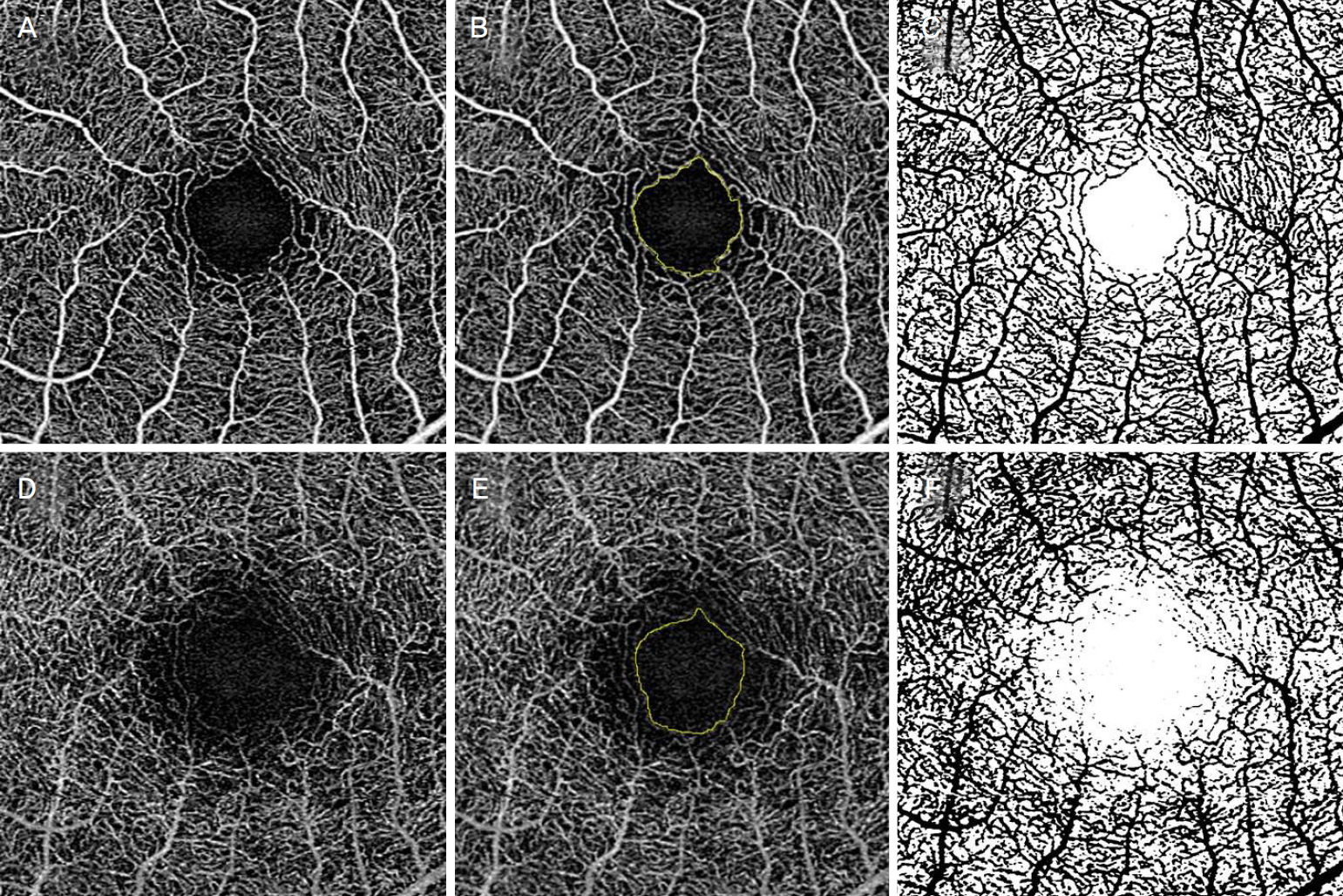
빛간섭단층혈관조영술 영상은 각 기기별로 기본으로 설정된 경계를 바탕으로 한 망막혈관의 표층모세혈관총과 심부모세혈관총 데이터를 획득하였다. 각 층의 망막혈관 영상을 Image J (Version 1.51, National Institutes of Health, Bethesda, Maryland, USA)를 사용하여 분석하였으며, 망막혈관 밀도는 2명의 판독자가 3 × 3 mm2 영역 중 망막중심오목무혈관 부위를 중심으로 2.7 × 2.7 mm2 만큼 잘라내어 8 bit로 변환한 뒤 Auto threshold with Otsu method를 이용하여 처리한 영상을 바탕으로 전체 영상의 혈관이 차지하는 면적을 계산하여 이를 비율로 계산하였다[14]. 망막중심오목무혈관부위는 2명의 판독자가 Image J를 이용하여 측정하였으며, 이의 평균값을 분석에 사용하였다(Fig. 1).
연구의 분석은, 통계학자의 조언에 따라 한 대상자에서 두 눈을 사용할 경우 발생할 수 있는 생물학적인 바이어스를 고려하여, 일괄적으로 좌안을 선택하여 분석하였다. 각 변수들은 Shapiro-Wilk 검정을 이용하여 표본의 정규성을 확인하였으며, 네 가지 기기에서 분석된 변수들의 평균은 Repeated Measure of analysis of variance를 사용하여 분석하였고, Bonferroni 교정을 이용한 사후검정을 시행하였다. 각 기기 간 변수의 상관성을 알아보기 위하여 Pearson 상관분석을 사용하였다. 통계분석에는 SPSS version 20.0 (IBM Corp., Armonk, NY, USA)을 사용하였고 p값이 0.05 미만인 경우 통계적으로 유의하다고 정의하였다.
결 과
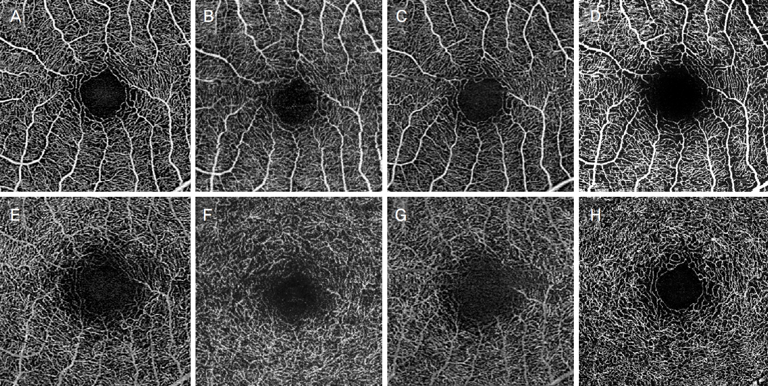
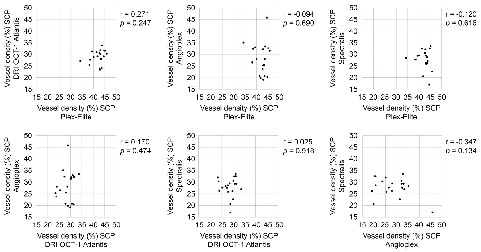
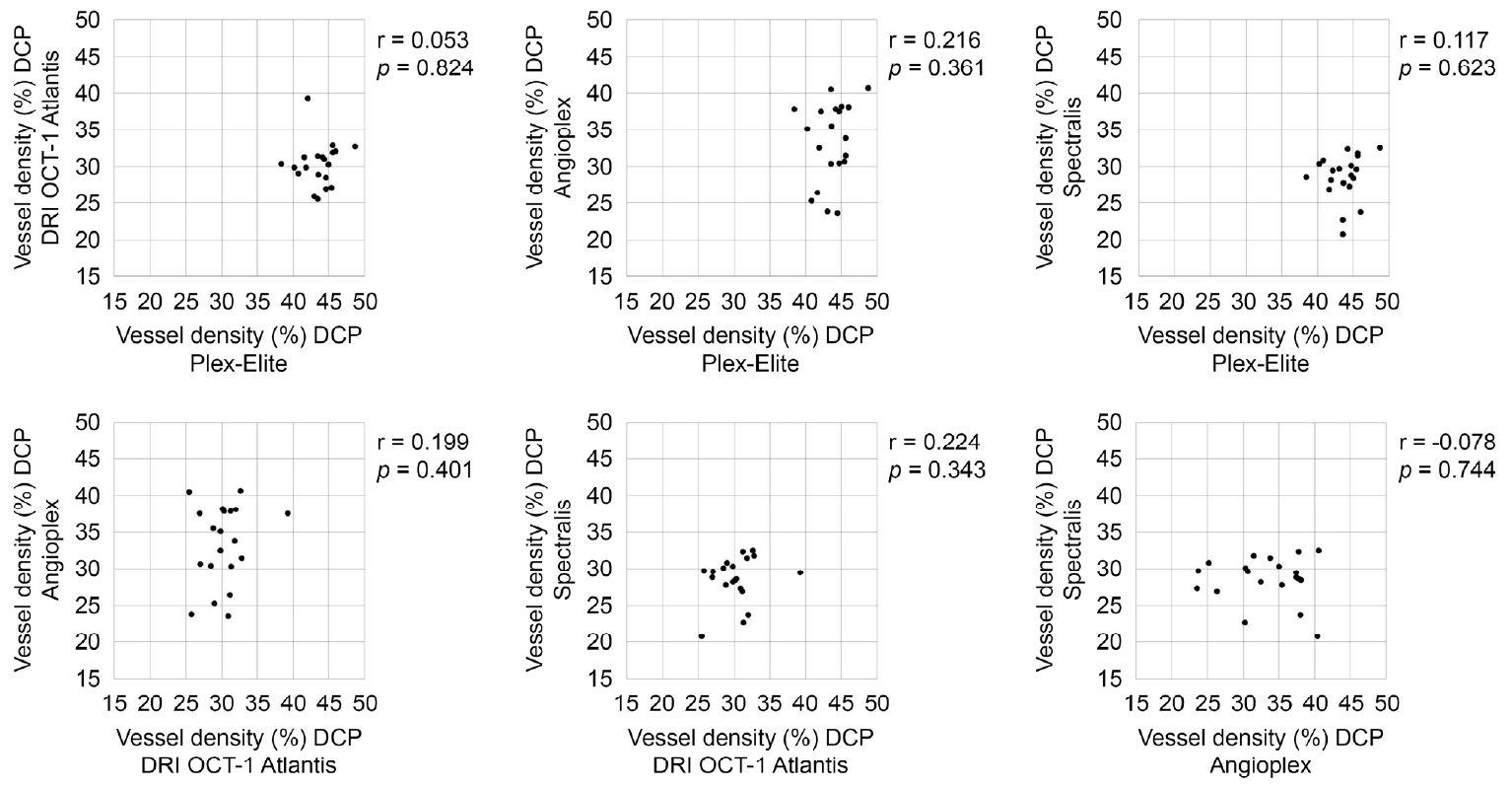
총 20명(20안)의 정상인이 포함되었으며, 평균 연령은 33.75 ± 4.04세(30-45), 남자는 10명(50%), 여자는 10명(50%)이 포함되었으며, 평균 안축장은 25.27 ± 0.62 mm였다. 망막혈관 밀도의 경우, 표층모세혈관총 및 심부모세혈관총 모두에서 네 가지 기기 간에 통계적으로 유의한 차이를 보였고(p<0.001), 사후 분석에서 두 층 모두에서 Plex-elite가 가장 높은 값을 나타냈다(p<0.001) (Table 2, Fig. 2). 표층모세혈관총 및 심부모세혈관총 망막혈관 밀도 모두 네 가지 기기 간 유의한 상관관계를 보이지 않았다(p>0.05) (Fig. 3, 4). 망막혈관 밀도의 관찰자 간 급내 상관계수는 표층 및 심부모세혈관총 각각 Plex-Elite (0.988, 0.963), DRI OCT-1 Atlantis (0.983, 0.976), AngioPlex (0.961, 0.966), Spectralis OCTA (0.968, 0.953)였다.
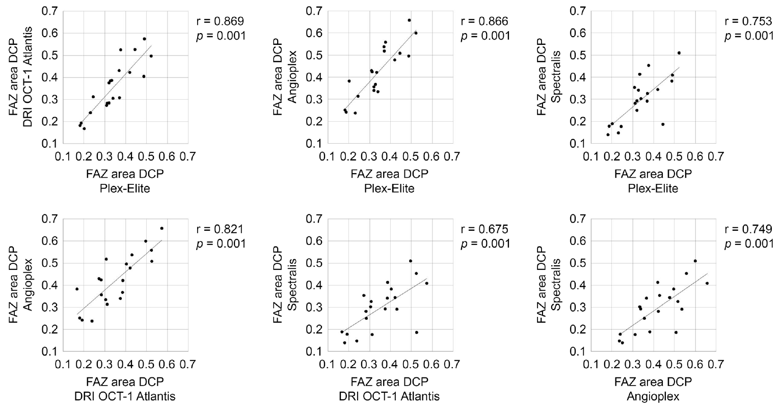
네 가지 기기 간의 망막중심오목무혈관부위 면적은 통계적으로 유의한 차이를 나타냈는데(p<0.001), 표층모세혈관총에서는 DRI OCT-1 Atlantis와 AngioPlex 간에 통계적으로 유의한 차이를 보였고(p=0.005), 심부모세혈관총에서는 Plex-Elite와 AngioPlex, DRI OCT-1 Atlantis와 AngioPlex, Spectralis와 AngioPlex 간에 통계적으로 유의한 차이를 보였다(p<0.001) (Table 2, Fig. 2). 망막중심오목무혈관부위 면적은 표층모세혈관총과 심부모세혈관총 모두에서 네 가지 기기 간의 통계적으로 유의한 양의 상관관계를 보였다(p<0.001) (Fig. 5, 6). 망막중심오목무혈관부위 면적의 관찰자 간 급내 상관계수는 표층 및 심부모세혈관총 각각 Plex-Elite (0.997, 0.961), DRI OCT-1 Atlantis (0.997, 0.992), AngioPlex (0.984, 0.981), Spectralis OCTA (0.981, 0.996)였다.
고 찰
본 연구는 최근 상용화된 빛간섭단층촬영기기 중 네 가지 빛간섭단층혈관조영술 기기(Plex-Elite, DRI OCT-1 Atlantis, AngioPlex, Spectralis OCTA)를 사용하여 망막혈관지표들을 분석하였다. 네 가지 기기를 이용하여, 정상인에서의 망막혈관 영상을 분석한 연구로, 동일한 개체에서 측정한 빛간섭단층혈관조영술 영상을 이용한 망막혈관 밀도 및 망막중심오목무혈관부위 영역은 기기 간의 차이가 있음을 알 수 있었다.
이번 연구에서 각 기기별로 측정한 표층 및 심부모세혈관총의 혈관 밀도는 각 기기별 차이가 있었으며, 사후분석상 Plex-Elite에서 가장 높은 값을 나타냈다. 빛간섭단층혈관조영술은 기본적으로 빛간섭단층촬영 영상에 기반한 것으로, 동일 구역에서 반복적인 촬영을 통해 얻어진 빛간섭단층촬영 영상을 이용한다[15,16]. 이러한 점을 고려하면, 빛간섭단층혈관조영술 영상을 획득하는 기기들 간의 특성 차이가 이러한 혈관 밀도의 차이를 나타낸 원인일 수 있다. 이는 기기별로 가지는 파장, 스캔 속도, 알고리즘 등의 특성에 기인할 수 있는데, 파장이 길수록 망막조직에 의한 산란이 적어져 좀 더 우수한 빛간섭단층혈관조영술 영상을 얻을 수 있는 것으로 알려져 있는 장점이 있다[17-19]. 하지만, 이러한 특징을 가지는 파장가변 빛간섭단층혈관조영술 기기의 경우 수평해상도가 스펙트럼영역 빛간섭단층혈관조영술 기기에 비해 떨어진다. 그러므로, 파장에 의한 해상도 차이가 영상의 질과도 연결될 수 있으며, 영상의 구현에 영향을 줄 수 있다. 또한 스캔 속도가 빠를수록 움직임 등에 의한 영상의 artifact를 줄일 수 있어, 더 좋은 질의 영상을 얻는 데 도움이 되나, 3 × 3 mm2 영역의 영상을 촬영하는데 큰 영향을 줄 것이라 판단하기는 어렵다. 빛간섭단층혈관조영술 기기의 알고리즘 중 OCT-based optical microangiography (OMAG)과 OCT angiography ratio analysis (OCTARA)는 motion detection을 위하여 intensity decorrelation과 phase variance 두 가지를 모두 사용하기 때문에 좋은 질의 영상을 얻는 데 유리하다고 알려져 있는데, 이번 연구에서 Plex-Elite는 깊은 파장과 빠른 스캔 속도, OMAG 알고리즘을 가지는 기기로서 망막혈관 밀도가 다른 기기보다 높게 측정되었을 가능성을 생각할 수 있다[3]. Spectralis OCTA는 full-spectrum amplitude decorrelation algorithm (FSADA) 알고리즘을 사용하므로 motion detection 시 amplitude decorrelation만을 사용하기 때문에 OMAG를 사용하는 AngioPlex와는 다른 결과를 나타낼 수 있다[3]. 추가적으로, 빛간섭단층혈관조영술 기기 내에 규정된 구획 경계가 기기 간에 다르므로 각 층의 망막혈관이 다르게 나타날 수 있음도 고려해야 하며, 또한 이러한 연구 결과를 해석함에 있어 주의해야 할 것은 영상을 처리하는 과정에서 선택하는 영상처리 방법에 따라, 정량적인 분석 결과가 달라질 수 있다는 점이다[20]. 그리고, 본 연구에 포함된 기기 중 AngioPlex만이 자동으로 표층모세혈관총의 망막혈관 밀도를 제공하기 때문에 영상 분석과정에서 모두 수동적으로 분석하였고, 이러한 과정이 연구 결과에 영향을 미쳤을 수 있다.
서로 다른 빛간섭단층혈관조영술 기기를 사용하여 망막혈관지표들을 분석한 이전의 연구 결과가 있었다. Munk et al [21]에 따르면, 건강한 성인에서 네 가지 서로 다른 빛간섭단층혈관조영술 기기(DRI-OCT Triton Swept-source OCT, prototype of Spectralis OCT2, AngioPlex, RTVue XR Avanti)로 측정한 망막혈관 밀도가 서로 통계적으로 유의한 차이를 보이지 않았다고 보고한 바 있다. 본 연구와 비교를 해보았을 때, 결과가 다른 것은 Plex-Elite 기기의 높은 망막혈관 밀도 때문인 것으로 판단되며, Munk의 연구에서 사용된 기기들 중 세 가지 기기(DRI-OCT Triton Swept-source OCT, prototype of Spectralis OCT2, AngioPlex)의 결과를 본다면, 본 연구의 결과는 Munk의 연구 결과와 유사하다고 판단된다. 하지만, 두 연구 모두 증례 수가 많지 않은 점을 고려하면, 더 많은 증례 수를 분석한 연구 결과가 필요하다고 판단된다. Corvi et al [13]은 일곱 가지 빛간섭단층혈관조 영술 기기(RTVue XR Avanti, prototype Spectralis, AngioPlex Cirrus 5000 HD, prototype Plex-Elite, RS-3000 Advance, OCT-HS100, Revo NX)에서 측정된 망막혈관 밀도는 상호 교환하여 사용할 수 없다고 보고하였다. 이는 본 연구와도 유사한 연구 결과이며, 이 연구에서도 다른 모든 기기들보다 Plex-Elite 기종의 망막혈관 밀도가 높았다. 이 연구에서 역시, 이러한 차이는 빛간섭단층촬영기기 간의 영상 구현의 방법, 알고리즘, 구획 경계(segmentation boundary)의 차이에 따른 결과일 것으로 설명하였다.
망막중심오목무혈관부위 면적은 표층모세혈관총보다 심부모세혈관총에서 더 큰 평균값을 보였고 각 기기들 간 차이가 있었다. 하지만 망막혈관 밀도와는 달리, 기기의 측정치 간 상관계수는 0.846에서 0.988까지 높은 값을 나타내어, 각 기기 간의 값의 차이는 있지만, 기기별로 측정한 값은 서로 양의 상관관계가 있음을 알 수 있었다. 이 중 표층모세혈관총의 망막중심오목무혈관부위의 기기 간의 상관계수는 0.846에서 0.988까지로, 심부모세혈관총의 망막중심오목무혈관부위의 기기 간의 상관계수 0.675에서 0.869 보다 높은 경향을 나타내었다. 이는 빛간섭단층혈관조영술을 이용한 심부모세혈관총의 망막중심오목무혈관부위의 경계가 표층모세혈관총보다 더 넓고 불분명하다고 보고한 이전의 연구를 바탕으로[22], 표층모세혈관총의 망막중심오목무혈관부위가 비교적 각 기기들 간의 일치도가 높다고 설명할 수 있다. 표층모세혈관총의 혈관 구조가 수평적으로 연결되어 있고 심부모세혈관총은 Vortex의 형태를 갖기 때문에 망막중심오목무혈관부위의 경계가 불분명하여 이러한 차이를 유발할 것이라 생각할 수 있고[23], 이는 두 검사자 간 일치도를 판단하는 표층모세혈관총의 급내 상관계수가 심부모세혈관총의 급내 상관계수보다 높았던 것을 고려하면, 이러한 차이를 보이는 이유를 설명할 수 있겠다.
본 연구는 상대적으로 대상자 수가 적은 편이며, 동일 대상에 대한 반복 측정을 하지 않아 검사 간 일치도를 분석하지 못한 한계점이 있다. 또한, 본 연구는 전향적으로 수행되었으나, 일부 증례에서 같은 시간, 같은 검사자에 의해 수행되지 못하여 이로 인해 발생할 수 있는 차이를 보정하지 못하였다. 망막혈관의 혈류는 일정하게 유지되고[24], 이를 촬영하는 빛간섭단층혈관조영술의 경우 검사 간 반복성이 높지만[25], 정상인을 대상으로 이에 대한 일중변동이나 검사 간 반복성에 대한 연구 결과가 없어, 이에 대한 보정이 필요할 수 있으며, 추후 이를 보완한 연구가 필요할 것으로 생각된다. 또한, 상대적으로 젊은 연령대의 대상자를 등록하여 수행한 연구로, 좁은 연령대의 데이터는 전체 연령 인구에서의 결과를 대표하지 못할 수도 있다. 또한, 혈압 등 혈류에 영향을 줄 수 있는 데이터를 수집하지 않아, 이의 영향을 분석하지 못하였는데, 이전 다른 연구에서는 망막혈류는 상대적으로 큰 변화 없이 유지되는 것으로 보고되고 있다[26].
결론적으로 네 가지 서로 다른 빛간섭단층혈관조영술 기기로 측정한 망막혈관 밀도와 망막중심오목무혈관부위 면적은 차이가 있었다. 망막혈관 밀도는 기기 간 상관 관계를 보이지 않았으며, 망막중심오목무혈관부위 면적은 기기 간 상관관계를 보였다. 이는 서로 다른 기기로 측정한 망막중심오목무혈관부위 면적과 망막혈관 밀도는 상호교환가능하지 않음을 시사하고, 빛간섭단층혈관조영술 영상을 분석하는 데 있어 이러한 차이를 고려해야 할 것으로 판단된다.


 PDF Links
 PubReader
 ePub Link
 Full text via DOI
 Download Citation
 Print
Print


