


개방각녹내장에서 해부학적 손상이 대개 기능 상실보다 선행한다고 알려져 있다[1]. 일부 연구에서 시야검사상 녹내장성 손상 발생 이전에 망막의 신경절세포와 그것들의 축삭돌기 손상이 발생한다고 보고한 바 있다[2,3]. 시신경유두나 망막신경섬유층 변화와 같은 해부학적인 손상 이후 기능적 손상이라고 여겨지는 표준 시야검사상의 시야손상이 나타난다[4]. 그러므로 초기 녹내장 진단 및 진행을 관찰하는 데 해부학적 손상을 관찰하는 것이 중요하다.
고도근시가 녹내장의 위험인자라는 것은 여러 연구에서 밝혀졌다[5,6]. 그러나 수직 타원형의 시신경유두, 시신경유두의 기울어짐, 이측 시신경테의 소실, 크고 얕은 함몰, 시신경유두주위 위축 등 고도근시의 이러한 시신경유두의 특징으로 인해[7], 고도근시에서 형태학적으로 녹내장의 조기 진단이 어려울 수 있다[8].
빛간섭단층촬영(optical coherence tomography, OCT) 장비가 발달하면서 시신경의 모양을 분석하고, 망막신경섬유층의 두께를 측정하여 녹내장을 진단하고 진행을 확인할 수 있다[9]. 최근 개발된 파장가변 빛간섭단층촬영(swept-source OCT, SS-OCT)는 1,050 nm의 중심파장과 약 100 nm의 대역폭을 가지는 파장 가변형 레이저를 광원으로 사용하는 장비로, 850 nm대의 파장 영역을 이용하는 기존의 스펙트럼영역 빛간섭단층촬영(spectral-domain OCT, SD-OCT)와 비교하여 더 긴 파장(더 높은 침투력)과 빠른 검사가 가능하다. 녹내장 진단에서 SS-OCT의 망막신경섬유층 두께 측정 정확도(100,000 to 400,000 A-scan/sec)가 기존 SD-OCT와 비슷한 것으로 보고되고 있다[10,11]. 시야손상 전 녹내장과 초기 녹내장에서 SD-OCT와 SS-OCT의 진단력을 비교한 연구에서 두 검사의 진단력은 비슷하다고 보고하였다[12]. 그러나 시신경 구조가 다른 고도근시 녹내장에서 두 가지 빛간섭단층촬영의 구조-기능 관계를 분석한 연구는 아직 드물다. 본 연구는 고도근시 개방각녹내장에서 스펙트럼 영역 빛간섭단층촬영과 파장가변 빛간섭단층촬영의 구조-기능 관계를 각각 분석해보고자 한다.
대상과 방법
2017년 9월부터 2019년 9월까지 서울성모병원 녹내장 클리닉을 방문한 환자 중 시야결손 전 개방각녹내장 및 원발개방각녹내장 환자를 대상으로 의무기록을 후향적으로 분석하였다. 본 연구는 헬싱키선언에 입각한 가톨릭대학교 의학연구윤리심의위원 회의 승인 아래 진행되었다(IRB 승인 번호: KC20RASI0001). 모든 환자들은 시력검사, 자동굴절검사, 골드만압평안압계검사, 세극등현미경검사, 전방각경검사, 안축장측정검사, 중심각막두께측정검사, 안저검사를 시행하였다. 전방각은 개방되어 있고, 전반적이거나 국소적인 시신경유두테의 좁아짐, 패임, 시신경유두출혈, 시야손상과 일치하는 망막신경섬유층 결손과 같은 특징적인 녹내장성 시신경손상이 있는 환자를 대상으로 하였다. 안내 수술(단순 백내장, 굴절교정수술 제외)을 받은 경우와 녹내장 이외의 시야 손상을 유발할 수 있는 망막전막, 당뇨망막병증, 망막혈관폐쇄 질환 등과 같은 망막 질환이 있는 경우는 제외하였다. 상기 환자 중 안축장길이 28 mm 이상의 고도근시안 43안을 대상군으로, 안축장길이 21 mm 이상 24 mm 미만인 30안을 대조군으로 선정하였다.
빛간섭단층촬영검사는 스펙트럼-영역 빛간섭단층검사기(CirrusTM HD-OCT ver. 6.0; Carl Zeiss Meditec, Inc., Dublin, CA, USA)와 파장가변 빛간섭단층검사기(DRI OCT TritonTM; Topcon Co., Tokyo, Japan)를 사용하였다. SD-OCT는 optic disc cube mode (200 × 200 scan)를 통해 망막신경섬유층 두께를 측정하였다. 시신경유두를 중심으로 직경 3.46 mm 부위의 자동으로 추출된 측정값을 사용하였다. 신호 강도가 6 미만이거나 중심에서 이탈된 영상은 제외하였다. SS-OCT는 초광각 스캔 프로토콜로 망막신경섬유층 두께를 측정하였다. 시신경유두를 중심으로 직경 3.4 mm 부위의 자동으로 추출된 측정값을 사용하였다. 영상의 질 점수 50 미만은 제외하였다. 두 검사에서 모두 눈의 움직임이나 측정의 부정확성으로 인하여 분석이 되지 않는 경우는 대상안에서 제외하였다. 빛간섭단층촬영검사로 측정한 시신경유두주변 망막신경섬유층 두께를 평균(360° 측정), 상비측(91°-135°), 비측(136°-225°), 하비측(226°-270°), 하이측(271°-315°), 이측(316°-45°), 상이측(46°-90°)으로 나누어 분석하였다.
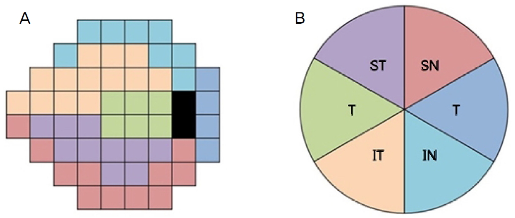
시야검사는 swedish interactive threshold algorithm (SITA) standard 24-2 방식으로 자동시야계(HumphreyTM, Carl Zeiss Meditec Inc.)로 시행하였다. 주시상실률, 위양성률, 위음성률 모두 15% 이상인 검사는 제외하였다. 시야검사의 mean deviation (MD), pattern standard deviation (PSD) 및 평균 시야감도와 영역별 시야감도를 분석하였다. 시야검사에서 데시벨(dB) 단위의 총 52군데의 역치값을 이용해, 평균 시야감도는 52군데 민감도의 평균으로 계산했다. 영역별 시야감도는 Garway-Heath et al [13]이 제시한 구조-기능 대응 지도에 따라 각 부분의 민감도를 계산하였다(Fig. 1). 생리적 맹점의 위, 아래쪽의 민감도는 제외하였다.
통계분석은 IBM SPSS ver. 22.0 software (IBM Corp., Armonk, NY, USA)를 사용하였다. 고도근시안과 비고도근시안군의 연령, 성별, 중심각막두께, 구면렌즈대응치, 안축장 길이, 시신경유두주위 위축 유무, 시야검사의 MD, PSD 비교 시 t-검정(student’s t-test)과 카이제곱검정(χ2 test)을 이용하였다. 두 가지 빛간섭단층촬영검사의 평균 및 사분면의 망막신경섬유층 두께 비교는 대응표본 t-검정(paired t-test)을 이용하였다. 망막신경섬유층 두께와 시야 민감도의 관계, 평균 망막신경섬유층 두께와 MD의 관계는 각각 단순선형회귀분석과 이차 다항회귀분석을 이용하여 분석하였다. SD-OCT와 SS-OCT의 구조-기능 관계를 비교하기 위해 Hotelling-Williams test를 이용하였다. p값이 0.05 미만인 경우를 통계학적으로 유의한 것으로 정의하였다.
결 과
개방각녹내장 환자 중 고도근시군 43안과 비고도근시군 30안을 포함하였다. 비고도근시군의 평균 연령은 67.8 고도근시군의 평균연령은 비고도근시군에 비해 유의하게 높았다(p=0.028). 개방각녹내장 중 시야손상 전 녹내장과 원발 개방각녹내장의 비율, 성별, 중심각막두께는 두 군 간의 유의한 차이가 없었다(p=0.965, p=0.375, p=0.751). 굴절력은 비고도근시군 -0.731.37 diopters (D), 고도근시군 -7.356.08 D로 고도근시군의 근시 정도가 유의하게 컸으며(p<0.001); 안축장 길이 비고도근시군 23,090.62 mm, 고도근시군 30,572.40 mm로 고도근시의 안구길이가 유의하게 길었다(p<0.001). 시신경유두주위 위축은 고도근시군이 42안, 비고도근시안이 10안으로, 고도근시안에서 유의하게 더 많았다(p<0.001). 시야검사상 MD는 두 군 사이 유의한 차이가 있었고, PSD는 비슷한 결과를 보였다(p=0.009, p=0.841). 평균 망막신경섬유층 두께를 SD-OCT로 측정하였을 때 두 군 사이 비슷한 결과를 보였고, SS-OCT로 측정하였을 때 고도근시안이 유의하게 얇게 측정되었다(p=0.35, p<0.001, Table 1).
고도근시안에서 SS-OCT로 망막신경섬유층 두께를 평균, 사분면으로 나누어 비교한 결과, SD-OCT와 비교하여 망막신경섬유층 두께가 유의하게 얇게 측정되었다(평균, 비측, 하측, 이측 p<0.001; 상측 p=0.008). 비고도근시안에서는 하측에서 두 가지 빛간섭단층촬영검사에서 비슷하게 측정되었고(p=0.053), 그 외 평균, 상측, 비측, 이측은 SD-OCT가 유의하게 얇게 측정되었다(p<0.001, Table 2).
고도근시군에서 SS-OCT로 측정한 평균 망막신경섬유층 두께와 시야검사에서의 평균 시야 감도는 일차 선형회귀, 이차 다항회귀분석에서 통계적으로 유의한 상관관계를 보였다(선형 r2=0.374, p<0.001; 이차 다항 r2=0.426, p<0.001). 시야검사의 MD과 평균 망막신경섬유층 두께는 일차 회귀, 이차 다항회귀분석 모두에서 유의한 관계를 보였다(선형 r2=0.362, p<0.001; 이차 다항 r2=0.409, p<0.001, Table 3). 망막신경섬유층을 여섯 개의 영역으로 나누어 각각에 대응하는 부분의 시야감도와의 관계를 일차회귀분석, 이차 다항회귀로 분석하였을 때, 망막신경섬유층과 시야감도는 6개의 모든 영역에서 유의한 상관관계를 보였다(모두 p<0.05, Table 4).
고도근시군에서 SD-OCT로 측정한 평균 망막신경섬유층 두께와 평균 시야 감도는 유의한 상관관계를 보이지 않았다(선형 r2=0.090, p=0.050; 이차 다항 r2=0.137, p=0.053). 평균 망막신경섬유층 두께와 시야검사의 MD을 분석했을 때 역시 유의한 상관관계를 보이지 않았다(선형 r2=0.085, p=0.059; 이차 다항 r2=0.126, p=0.067; Table 3). 영역별 망막신경섬유층 두께와 대응하는 시야 감도를 분석했을 때, 일차 선형분석에서는 하이측에서 유의한 상관관계를 보였으나(r2=0.127, p=0.019), 이차 다항함수에서는 상관관계를 보이지 않았다(r2=0.137, p=0.053). 이차 다항회귀분석 시상이측(r2=0.296, p<0.001)과 상비측(r2=0.151, p=0.038)에서 유의한 상관관계를 보였으나, 일차 선형회귀분석 시 유의한 상관 관계를 보이지 않았다(상이측 r2=0.083, p=0.061; 상비측 r2=0.025, p=0.308; Table 4).
비고도근시안에서 SS-OCT로 측정한 평균 망막신경섬유층 두께와 평균 시야감도는 통계적으로 유의한 상관관계를 보였다(선형 r2=0.371, p<0.001; 이차 다항 r2=0.411, p<0.001). 시야검사의 MD와 평균 망막신경섬유층 두께 또한 유의한 관계를 보였다(선형 r2=0.424, p<0.001; 이차 다항 r2=0.487, p<0.001; Table 3). 상이측, 하이측, 상비측, 하비측에서 각각의 망막신경섬유층과 시야검사 민감도는 통계적으로 유의한 상관관계를 보였다(모두 p<0.05, Table 4).
비고도근시군에서 SD-OCT로 측정한 평균 망막신경섬유층 두께와 평균 시야감도 사이에는 유의한 상관관계를 보이지 않았다(선형 r2=0.116, p=0.066; 이차 다항 r2=0.374, p=0.130). 그러나 시야검사의 MD와 평균 망막신경섬유층 두께를 비교했을 때, 일차 선형회귀분석에서는 통계적으로 유의한 상관관계를 보였고(r2=0.148, p=0.036), 비선형분석에서는 유의한 관계를 보이지 않았다(r2=0.159, p=0.097; Table 3). 상이측, 하이측, 상비측, 하비측에서 망막신경섬유층 두께와 대응하는 시야검사의 민감도는 유의한 상관관계를 보였다(모두 p<0.05, Table 4).
Hotelling-Williams test를 이용하여 SD-OCT와 SS-OCT의 구조-기능 관계를 통계적으로 비교하였다. 고도근시군에서 SD-OCT와 SS-OCT의 평균 망막신경섬유층 두께와 시야검사 MD, 그리고 평균 시야감도 사이 관계는 각각 일차 선형회귀분석에서 통계적으로 유의한 차이를 보였다(시야검사 MD p=0.047; 평균 시야감도 p=0.040, Table 3). 고도근시군에서 일차 선형회귀분석 시, 이측, 상비측, 하비측에서, 이차 다항회귀분석 시, 이측, 상비측에서 두 OCT의 구조-기능 상관관계가 통계적으로 유의한 차이를 보였다(모두 p<0.05, Table 4). 비고도근시군에서 일차 선형분석과 이차 다항분석을 이용한 두 가지 OCT의 평균 망막신경섬유층 두께와 시야검사 MD, 그리고 평균 망막신경섬유층 두께와 평균 시야감도의 상관관계는 각각 유의한 차이를 보였다(모두 p<0.05, Table 3). 비고도근시군에서 일차 선형회귀분석 시, 하이측, 이측, 비측에서, 이차 다항회귀분석 시, 하이측, 이측에서 두 OCT의 구조-기능 관계가 통계적으로 유의한 차이를 보였다(모두 p<0.05, Table 4).
고 찰
정시안에 비해 근시안의 빛간섭단층촬영으로 측정한 망막신경섬유층 두께는 차이가 있다. 이전 연구에서 근시안은 정시안에 비해 평균 시신경유두 주위 망막신경섬유층 두께가 얇다고 밝혀졌다[14-16]. 근시에서는 망막신경섬유층 두께의 패턴이 달라지는데, 상이측, 하이측의 망막신경섬유들이 이측으로 집중되는 경향이 있다[17-19]. Fang et al [20]은 중등도(-3.00 이하 -6.00 D 초과) 및 고도근시(-6.00 D 이하)에서 SD-OCT가 녹내장 진단 특이도가 낮다고 보고하고 있다. 고도근시는 형태학적으로 정시안과 다르기 때문에 고도근시 녹내장은 진단에 어려움이 있다.
Kerrigan-Baumrind et al [21]가 적어도 25%에서 35%의 망막신경절세포의 손실이 시야검사상 손상과 연관되어 있다고 발표한 이후로 녹내장에서 시야결손 전 망막신경절 세포 손상이 선행한다고 알려져 있다. 구조와 기능 검사를 결합하면 녹내장의 진단력을 높일 수 있다[22]. 빛간섭단층검사가 time-domain OCT에서 SD-OCT, 그리고 SS-OCT로 발전되면서 각각의 OCT에 대한 구조-기능관계를 분석하는 많은 논문이 발표되었다. Hood and Kardon [1]와 Leite et al [23]의 연구에서 자동시야검사와 빛간섭단층촬영의 망막신경섬유층 두께의 구조-기능 선형의 구조 관계를 규명하였다. SD-OCT의 시신경유두주변 망막신경섬유층두께나, 시신경유두테의 폭 등 빛간섭단층촬영의 여러 계측치를 분석하여 구조-기능 관계를 밝히는 연구가 보고되었고[24,25], 시야결손 전 녹내장에 비해 시야결손이 나타난 녹내장에서 우월한 구조-기능 관계를 보인다고 밝힌 바 있다[26,27]. 그리고 Raza and Hood [28]은 녹내장에서 SS-OCT의 망막신경절세포층 두께와 시야 감도가 유의한 상관관계를 보인다고 보고하였다. 하지만 아직 고도근시 녹내장에서 SD-OCT나 SS-OCT의 구조-기능 관계에 대해 발표한 논문은 없다.
지금까지 많은 연구에서 녹내장 및 정상안에서 SD-OCT와 SS-OCT를 비교해왔다. Lee et al [29]은 정상안에서 SD-OCT와 SS-OCT의 망막신경섬유층 두께 및 신경절세포 두께 측정의 정확도는 비슷하다고 보고하였다. Lee et al [12]은 시야손상 전 녹내장과 초기 녹내장에서 SD-OCT와 SS-OCT의 망막신경섬유층 두께 지도의 진단 정확도는 비슷하다고 보고하였다. Lee et al [12]의 연구는 초광각 SS-OCT와 SD-OCT인 Cirrus HD-OCT로 망막신경섬유층 두께를 측정하였다는 점에서 본 연구와 비슷하다. 하지만 본 연구와 차이점이 있다. 첫째, 구면렌즈대응치 +6.0에서 -6.0 D인 환자군을 대상으로 했으므로 고도근시안을 포함하지 않았다. 둘째, 두 가지 OCT에서 망막신경섬유층 두께 지도를 측정하여 정성적 분석만 시행하였다. 본 연구는 망막신경섬유층 두께의 평균 및 영역별 값을 통해 정량적 분석을 시행하였다.
Ha et al [30]은 녹내장, 녹내장 의증, 건강안에서 SS-OCT가 SD-OCT보다 망막신경섬유층이 유의하게 두껍게 측정된다고 보고하였다. Lee et al [12]도 시야손상 전 녹내장과 초기 녹내장에서 SS-OCT의 망막신경섬유층 두께가 SD-OCT보다 두껍게 측정된다고 보고하였다. 본 연구에서도 비고도근시군에서는 SD-OCT에 비해 SS-OCT의 평균과 하측을 제외한 3개의 영역의 망막신경섬유층 두께가 유의하게 두껍게 측정되었다(p<0.001). 이는 이전의 연구와 일치하는 결과이다. SS-OCT에서 스캔하는 원의 반지름(3.40 mm)이 SD-OCT (3.46 mm)에 비해 작기 때문이라고 추정하고 있다[12]. 본 연구에서 SD-OCT와 SS-OCT의 스캔하는 원의 반지름은 각각 3.46 mm, 3.40 mm로 기존의 연구와 동일하다. 망막신경섬유층은 시신경유두 주변에서 두껍고, 주변부에서 더 얇기 때문에[31], 스캔한 원이 작을수록 망막신경섬유층 두께가 두껍게 측정될 수 있기 때문이다. 하지만 아직까지 두 OCT의 망막신경섬유층 두께의 차이에 대한 명확한 원인은 밝혀지지 않았다. 이와 상반되어 고도근시군에서는 SD-OCT에 비해 SS-OCT에서 평균 및 상측을 제외한 사분면의 망막신경섬유층 두께가 유의하게 얇게 측정되었다(p<0.001). 지금까지 고도근시 녹내장에서 SS-OCT와 SD-OCT의 진단력을 비교한 연구는 발표되지 않았다. 대부분의 정시안 녹내장의 연구에서는 본 연구와 마찬가지로 SD-OCT에 비해 SS-OCT의 망막신경섬유층 두께가 두껍게 측정되었다. 기존 SD-OCT는 시신경유두에 초점을 맞추어 영상을 추출하는 반면, 본 연구에서 시행한 초광각 SS-OCT는 시신경유두와 황반 사이의 영역에 중심을 두어 영상을 추출한다[32]. 고도근시는 수직 타원형의 시신경유두, 시신경유두의 기울어짐, 시신경유두주위 위축 등 시신경유두의 형태학적 왜곡이 있다. SS-OCT와 SD-OCT의 영상을 스캔 방식의 차이로 인해 SS-OCT가 고도근시의 왜곡된 시신경 형태를 더 잘 반영한다고 추정된다. 앞으로 고도근시 녹내장에서 SS-OCT의 망막신경섬유층 및 신경절세포층 두께 등 진단 정확도에 대한 더 많은 연구가 필요할 것이다.
본 연구에서 비고도근시군에서 평균 망막신경섬유층 두께와 시야검사상 평균감도는 SD-OCT에서 유의한 상관관계를 보이지 않았고(p>0.05), SS-OCT에서는 통계적으로 유의한 상관관계를 보였다(p<0.05). 시야검사의 MD과 평균 망막신경섬유층 두께의 상관관계를 분석하였을 때, SS-OCT의 경우 일차선형, 이차다항회귀분석에서 모두 유의한 상관 관계를 보였으나(p<0.001), SD-OCT의 경우 일차선형회귀분석에서는 유의한 결과를(p=0.036), 이차다항회귀분석에서는 유의하지 않은 결과를 보였다(p=0.097). 지금까지 여러 연구에서 녹내장에서 시야검사의 MD과 SD-OCT의 평균 망막신경섬유층 두께는 유의한 상관관계를 보인다고 보고했다[33,34]. 본 연구에서 비고도근시군의 SD-OCT의 경우 MD과 평균 망막신경섬유층 두께의 선형 회귀 분석에서 유의한 결과를 보였으며, 이는 이전 보고된 연구들과 동일한 결과이다. 두 가지 OCT에서 모두 이측과 비측을 제외한 상이측, 하이측, 상비측, 하비측에서 유의한 상관관계를 보였다(p<0.05). 한 연구에서 SD-OCT의 비측과 이측의 망막 신경섬유층 두께가 해당하는 시야검사와 낮은 상관관계를 보인다고 보고했다[35]. 그러므로 비고도근시에서는 SD-OCT와 SS-OCT는 각각 평균 및 영역별 구조-기능 간 유의한 상관관계를 보인다.
고도근시군에서 평균 망막신경섬유층 두께와 전체 시야감도와 MD은 SD-OCT 경우 유의한 상관관계를 보이지 않았고(p≥0.05), 각 영역별 선형회귀분석으로는 하이측에서 상관관계를 보였으나(p=0.019) 이차 다항회귀분석상에서는 상이측, 상비측에서 상관관계를 보였다(p=0.001, p=0.038). SD-OCT는 고도근시 녹내장에서 구조-기능 상관관계가 뚜렷하지 않았다. 하지만 SS-OCT상 평균 망막신경섬유층 두께와 전체 시야검사 및 6개의 모든 영역의 망막신경섬유층 두께와 그에 대응하는 시야검사상 매우 높은 상관관계를 보였다(p<0.05). Hotelling-Williams test를 이용하여 두 OCT 간 구조-기능 상관관계를 비교하였다. 고도근시군에서 평균 시야감도, MD과 각각 평균 망막신경섬유층 두께의 상관관계를 비교하였을 때, 일차 선형회귀분석에서 유의한 차이를 보였다(p<0.05, Table 3). 일차 선형회귀분석 시, 이측, 상비측, 하비측에서, 이차 다항회귀분석 시, 이측, 상비측에서 두 OCT의 구조-기능 상관관계가 통계적으로 유의한 차이를 보였다(p<0.05, Table 4). 고도근시 녹내장에서 SD-OCT에 비해 SS-OCT가 상대적으로 뚜렷한 평균 및 영역별 구조-기능 관계를 보여주었다.
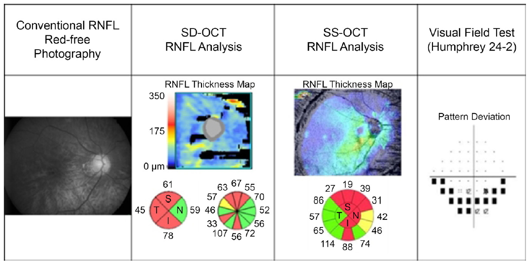
Fig. 2는 안축장 길이 30.99 mm인 고도근시 녹내장 환자로 SS-OCT으로 측정한 평균 망막신경섬유층 두께(58 μm)가 SD-OCT로 측정한 평균 망막신경섬유층 두께(61 μm)보다 얇게 측정되었다. SD-OCT는 망막신경섬유층 두께가 전반적으로 얇게 측정된 반면, SS-OCT는 시야손상과 일치하는 영역에 두드러지게 망막신경섬유층 두께가 얇게 측정되었다. 고도근시에서 SS-OCT가 SD-OCT에 비해 상대적으로 구조-기능 관계를 잘 보여주고 있다.
본 연구는 몇 가지 제한점을 가지고 있다. 첫째, 대상 환자 수가 상대적으로 적다. 그러나 고도근시안에서 각기 다른 종류의 빛간섭단층촬영의 구조-기능 관계를 밝힌 첫 연구라는 점에서 의미가 있다. 둘째, 본 연구는 Garway-Heath et al [13]이 제시한 구조-기능 대응 관계 지도를 통해 분석하였기에 시신경유두주위 위축 등 구조적으로 다른 고도근시안의 경우 결과가 다르게 나올 수 있다. 하지만 각 영역별로 나누어 분석했을 때, 고도근시군에서 SS-OCT의 경우 구조-기능 관련성이 유의하였고, SD-OCT의 경우 구조-기능 관련성이 유의하지 않았다는 점에서 그 오차는 낮을 것으로 사료된다. 추후 고도근시안에 대한 다른 구조-기능 모델에 대한 추가적인 연구가 필요할 것으로 생각된다. 셋째, 고도근시군에서 SS-OCT가 6개의 영역의 망막신경섬유층 두께와 그에 대응하는 시야검사상 높은 상관관계를 보였으나, 두 OCT의 상관관계의 차이는 일차 선형회귀분석 시 이측, 상비측, 하비측에서, 이차 다항 회귀 분석 시 이측, 상비측에서만 나타났다. 그러나 두 기기의 평균 망막신경섬유층 두께와 평균 시야감도, MD는 각각 유의한 상관관계 차이를 나타냈고, 이측, 상비측, 하비측에서 유의한 상관관계 차이를 보였다는 점에서 본 연구의 의미가 있다. 앞으로 고도근시에서 SD-OCT와 SS-OCT의 구조-기능 관계를 비교하는 대규모 연구가 필요할 것이다.
녹내장에서 시야결손이 발현되기 전에 구조적 변화를 감지하면 조기에 진단할 수 있어 이는 예후에 중요하다. 하지만 지금까지 고도근시 녹내장은 형태학적으로 달라 진단이 어려웠다. 본 연구는 고도근시 녹내장에서 SS-OCT인 Topcon DRI OCT가 SD-OCT인 Cirrus HD-OCT와 비교하여 전반적 및 국소적으로 구조-기능 관련성이 더 뚜렷하다는 것을 밝혔다. 고도근시 녹내장에서 SS-OCT를 진단적 기준으로 삼고 활용할 수 있는 근거가 될 수 있을 것으로 생각된다. 앞으로 고도근시 녹내장 환자에서 다양한 진단 도구에 대한 구조-기능 관계를 분석하여, 녹내장을 조기 진단하는 데 도움을 주어야 할 것이다.

 PDF Links
 PubReader
 ePub Link
 Full text via DOI
 Download Citation
 Print
Print


